
磁共振质子密度脂肪分数(MRI-PDFF)评估慢性HBV感染者肝脂肪变性及分布特点
2024-06-06 09:41王丽旻包超赵凯越靳婕华郑卓肇黄缘
临床肝胆病杂志 2024年5期
王丽旻 包超 赵凯越 靳婕华 郑卓肇 黄缘


摘要: 目的 利用磁共振質子密度脂肪分数 (MRI-PDFF) 评估慢性HBV感染者的肝脂肪变性的程度。方法 选择北京清华长庚医院2018年1月—2022年12月门诊或者住院, 明确诊断为慢性HBV感染, 年龄>16岁的患者。所有患者均在本院行肝脏磁共振检查。依据是否合并肝硬化进行分组, 比较患者的各个肝段PDFF值的一致性。使用Kappa一致性检验、 组内相关系数 (ICC) 进行一致性分析。结果 纳入76例核苷 (酸) 类似物经治患者, 其中30. 26% (23例) 合并肝硬化, 所有患者的MRI-PDFF算术均值波动在1. 49%~30. 93%, 依据MRI-PDFF≥5%诊断为脂肪肝, 38. 16% (29例) 患者合并脂肪肝。76例患者的全肝PDFF算术均值低于全肝PDFF加权均值, 全肝PDFF加权均值、 全肝PDFF算术均值、 左半肝及右半肝的PDFF算术均值比较, 差异无统计学意义 (F=0. 39, P=0. 76)。各个肝段、 左/右半肝PDFF算术均值与全肝PDFF加权均值、 全肝PDFF算术均值的一致性较强 (ICC均>0. 75), 但右半肝PDFF算术均值与全肝PDFF加权均值的一致性高于左半肝。合并肝硬化组患者在区分脂肪肝诊断的一致性检验方面, Ⅶ段PDFF算术均值与全肝PDFF加权均值一致性差 (Kappa=0. 39), 左半肝、Ⅰ、 Ⅱ、 Ⅲ、 Ⅴ、 Ⅵ、 Ⅷ为中等一致。合并肝硬化的患者Ⅶ段PDFF算术均值与全肝PDFF加权均值一致性最低, Ⅳ段一致性最高。未合并肝硬化的患者Ⅱ段PDFF算术均值与全肝PDFF加权均值一致性最低, Ⅴ段的一致性最高。结论 MRI-PDFF评估慢性HBV感染者的肝脂肪变性更全面, 合并肝硬化的患者各肝段PDFF算术均值与全肝PDFF加权均值的一致性差异大。
关键词: 乙型肝炎病毒; 脂肪肝; 磁共振成像
基金项目: 北京市自然科学基金 (7202240); 清华大学自主科研项目 (2022PY002)
Value of magnetic resonance imaging-proton density fat fraction in evaluating the degree and distribution
characteristics of hepatic steatosis in patients with chronic hepatitis B virus infection
WANG Limina, BAO Chaob, ZHAO Kaiyuea, JIN Jiehuaa, ZHENG Zhuozhaob, HUANG Yuana. (a. Department of Gastroenterology,
b. Department of Radiological Diagnosis, Beijing Tsinghua Changgung Hospital, Tsinghua University, Beijing 102218, China)
Corresponding author: HUANG Yuan, huangy9815@163.com (ORCID: 0000-0003-1392-4619)
Abstract:
ObjectiveTo investigate the value of magnetic resonance imaging-proton density fat fraction (MRI-PDFF) in evaluating hepatic steatosis in patients with chronic hepatitis B virus (HBV) infection. Methods The patients, aged >16 years, who visited the outpatient service or were hospitalized in Beijing Tsinghua Changgung Hospital from January 2018 to December 2022 and were diagnosed with chronic HBV infection were enrolled, and all patients underwent MRI examination of the liver in our hospital. The patients were divided into groups based on the presence or absence of liver cirrhosis, and the consistency in PDFF between different hepatic segments was compared between groups. The Kappa consistency test and intraclass correlation coefficient (ICC) were used for consistency analysis. Results A total of 76 patients treated with nucleoside analogues were enrolled, among whom 23 (30.26%) had liver cirrhosis. For all patients, the simple arithmetic average of PDFF fluctuated between 1.49% and 30.93%. According to MRI-PDFF ≥5% as the diagnostic criterion for fatty liver disease, there were 29 patients (38.16%) with fatty liver disease among all patients. For all 76 patients, the simple arithmetic average of PDFF was lower than the weighted average of PDFF for the whole liver, and there was no significant difference between the simple arithmetic average of PDFF, the weighted average of PDFF, and the PDFF values of the left and right lobes of the liver (F=0.39, P=0.76) . The consistency test showed that the PDFF values of each hepatic segment and the left and right lobes of the liver had strong consistency with the weighted average and simple arithmetic average of PDFF, with an ICC of >0.75, but the consistency between the PDFF value of the right lobe and the weighted average of PDFF was higher than that between the PDFF value of the left lobe and the weighted average of PDFF. In the consistency test of differentiating fatty liver disease in patients with liver cirrhosis, there was poor consistency between the PDFF value of segment Ⅶ and the weighted average of PDFF (Kappa=0.39), with moderate consistency for the left lobe and the Ⅰ, Ⅱ, Ⅲ, Ⅴ, Ⅵ, and Ⅷ segments. For the patients with liver cirrhosis, the lowest consistency was observed between the PDFF value of Ⅶ segment and the weighted average of PDFF for the whole liver, and the highest consistency was observed between the PDFF value of Ⅵ segment and the weighted average of PDFF for the whole liver. For the patients without liver cirrhosis, the lowest consistency was observed between the PDFF value of Ⅱ segment and the weighted average of PDFF for the whole liver, and the highest consistency was observed between the PDFF value of Ⅴ segment and the weighted average of PDFF for the whole liver. Conclusion MRI-PDFF is more comprehensive in evaluating hepatic steatosis in patients with chronic HBV infection, and for the patients with liver cirrhosis, there is poor consistency between the PDFF value of each segment and the weighted average of PDFF.
Key words: Hepatitis B virus; Fatty Liver; Magnetic Resonance Imaging
Research funding: Beijing Natural Science Foundation (7202240); Tsinghua University Independent Research (2022PY002)
慢性HBV感染者中合并肝脂肪变性的人数越来越多[1] , 而肝脂肪变性可能加速慢性肝病患者的肝纤维化进程[2] , 代谢性疾病包括代谢相关的非酒精性脂肪性肝病 (NAFLD) 可增加慢性HBV感染者的全因死亡率[3]。因此对于合并脂肪肝的患者, 除积极抗病毒治疗外, 筛查和管理代谢异常包括脂肪肝对于预防疾病进展有重要意义。磁共振成像 (MRI) 测量的肝脏质子密度脂肪分数 (proton density fat fraction, PDFF) 是当前无创性检测肝脂肪变性的 “金标准”, 与磁共振波谱和肝穿刺活检均有良好的相关性[4-6] 。本研究以MRI-PDFF检测慢性HBV感染者的肝脂肪变性程度, 评估其肝脂肪变性的分布特点及与疾病进程的关系。
1 资料与方法
1. 1 研究对象 选择2018年1月—2022年12月在本院就诊、 明确诊断为慢性HBV感染、 年龄>16岁、 病程>6个月的患者。所有患者均在本院接受过MRI检查。除外合并酒精性肝病、 自身免疫性肝病、 慢性丙型肝炎、 肝癌、 肝移植患者。肝硬化的诊断依据 《慢性乙型肝炎防治指南 (2022年版)》 [7] 。
1. 2 肝脏MRI检查 所有患者应用3. 0T扫描仪 (Discovery MR750, GE Healthcare, Waukesha, WI, USA)进行肝脏MRI检查。检查前患者禁食8 h, 检查时仰卧位, 使用腹部相控阵线圈, 常规扫描序列包括: 横断面呼吸门控脂肪抑制FSE T2WI、 横断面呼吸门控DWI和横断面呼气末屏气LAVA-Flex等序列。肝脏PDFF测量则采用呼气末屏气三维IDEAL-IQ序列, 序列参数如下: TR 6. 2 ms, 扫描野38 cm, 层厚8 mm, 翻转角3°, 一次三维采集时间为19 s。检查完成后, 在横断面肝脏脂肪分量图中勾画感兴趣区。依据Couinaud肝脏分段法, 肝脏分为肝段Ⅰ、 Ⅱ、Ⅲ、 Ⅳ、 Ⅴ、 Ⅵ、 Ⅶ、 Ⅷ, 在每个肝段勾画圆形感兴趣区, 直径约1 cm, 避开大胆管、 大血管、 病灶、 肝脏包膜、 肝裂及胆囊, 从而得到肝脏各段的 PDFF 值, 每个肝段均测3次, 取平均值作为该患者各段最终的PDFF值[6] 。
1. 3 统计学方法 采用SPSS 21. 0统计学软件包进行分析, 满足正态分布的计量资料以x ˉ±s表述, 非正态分布的计量资料以M (P25~P75 ) 表示。使用Kappa一致性检验、 组内相关系数 (intraclass correlation coefficient, ICC) 进行一致性分析。Kappa和ICC均<0. 40提示一致性較差, ≥0. 40~0. 75提示一致性中等, ≥0. 75提示一致性强。P<0. 05为差异有统计学意义。
2 结果
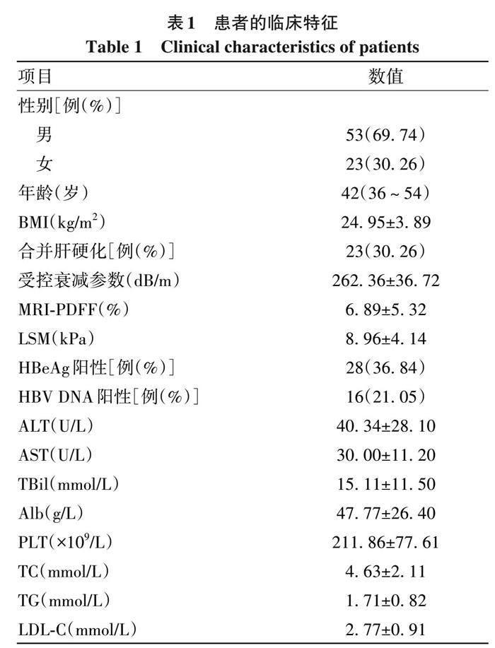
2. 1 患者的基本情况 本研究共纳入76例患者, 其中男53例 (69. 74%), 女23例 (30. 26%), 年龄16~80岁。患者均为核苷 (酸) 类似物经治 (包括恩替卡韦、 替诺福韦、 丙酚替诺福韦), 其中HBeAg阳性28例 (36. 84%),HBV DNA>30 IU/mL 16例 (21. 05%)。76例患者中23例(30. 26%) 合并肝硬化。所纳入患者的PDFF算术均值波动在 1. 49%~30. 93%, 中位数 4. 36%。依据 PDFF≥5%诊断为脂肪肝[8] , 合并脂肪肝患者29例 (38. 16%)。BMI≥24 kg/m2的超重患者44例 (57. 89%), ≥28 kg/m2的肥胖患者13例 (17. 11%)(表1) 。
2. 2 患者各肝段测量的PDFF 根据Couinaud肝段划分法, 肝段Ⅰ~Ⅳ属于左半肝, 肝段Ⅴ~Ⅷ属于右半肝。计算76例患者每个肝段、 全肝以及左半肝、 右半肝的PDFF算术均值。另外依据Mise等[9]的研究, 肝段Ⅰ、Ⅱ、 Ⅲ、 Ⅳ、 Ⅴ、 Ⅵ、 Ⅶ、 Ⅷ分别占全肝体积的4. 0%、 7. 9%、9. 5%、 13. 6%、 12. 6%、 7. 9%、 16. 8%、 26. 1%。分别对8个肝段进行加权计算, 获得全肝PDFF加权均值。对不同的PDFF值进行比较, 结果显示右半肝、 左半肝的PDFF算术均值, 全肝PDFF算术均值, 全肝PDFF加权均值比较, 差异无统计学意义 (F=0. 49, P=0. 76)。左半肝、 全肝的PDFF算术均值低于全肝PDFF加权均值, 右半肝相比左半肝PDFF算术均值与全肝PDFF加权均值更相近。Ⅷ肝段的PDFF算术均值最高, Ⅲ肝段最低 (表2) 。2. 3 PDFF评估肝脂肪变性程度的一致性分析 ICC分析结果提示, 各个肝段、 左/右半肝PDFF算术均值与全肝PDFF加权均值、 全肝PDFF算术均值的一致性较强, ICC均>0. 75, 右半肝PDFF算术均值与全肝PDFF加权均值的一致性高于左半肝。在所有肝段中, 肝Ⅴ段PDFF算术均值与全肝 PDFF 加权均值的一致性最高(ICC=0. 99), 肝Ⅳ、 Ⅶ段其次 (ICC=0. 98), 肝Ⅱ段最低 (ICC=0. 92)。全肝PDFF算术均值与肝Ⅲ段的一致性最低(ICC=0. 91)(表3)。
Kappa一致性检验结果显示, 区分PDFF≥5%和<5%时, 全肝 PDFF算术均值、 左半肝/右半肝及各肝段的PDFF算术均值与全肝PDFF加权均值一致性强 (Kappa均>0. 75); 右半肝PDFF算术均值与全肝PDFF加权均值的一致性高于左半肝 (0. 94 vs 0. 86); 各肝段中Ⅳ段和Ⅴ段的PDFF算术均值与全肝PDFF加权均值诊断脂肪肝的一致性最高 (Kappa=0. 91)(表4) 。
2. 4 依据是否合并肝硬化分组比较PDFF 本组患者中23例 (30. 26%) 合并肝硬化, 肝硬化组PDFF≥5%的患者11例 (47. 83%), 未合并肝硬化PDFF≥5%的患者18例(33. 96%)。依据合并肝硬化与否分组, 分别以全肝PDFF加权均值与左半肝、 右半肝、 各个肝段的PDFF算术均值做一致性检验 (表5)。结果发现合并肝硬化患者的ICC和Kappa值均明显低于未合并肝硬化组, 合并肝硬化的患者Ⅵ段PDFF算术均值与全肝PDFF加权均值的ICC为0. 74, 为中等一致。在合并肝硬化患者中, 区分脂肪肝 (PDFF≥5%) 诊断的一致性检验显示, Ⅶ段PDFF算术均值与全肝PDFF加权均值一致性差 (Kappa=0. 39), 左半肝、 Ⅰ、 Ⅱ、 Ⅲ、 Ⅴ、 Ⅵ、 Ⅷ为中等一致, 仅右半肝、 Ⅳ段的一致性强。合并肝硬化的患者Ⅶ段PDFF算术均值与全肝PDFF加权均值一致性最低, Ⅳ段最高。在未合并肝硬化的患者, 各肝段中Ⅱ段PDFF算术均值与全肝PDFF加权均值的一致性最低, Ⅴ段最高。
3 讨论
在隐源性原发性肝癌者中NAFLD是最常见的病因[10]。非酒精性脂肪性肝炎(NASH)患者肝细胞癌(HCC) 发病人数高达5. 29/1 000人年[11] 。慢性HBV感染者合并肝细胞脂肪变患病率达32. 8%[3] 。因此提高对慢性HBV感染者肝脂肪变性的诊断, 有利于对疾病的早干预。MRI-PDFF可测量肝脏的TC含量, 其敏感度和特异度均较高, 而且有较好的可重复性和再现性[12-13] 。与组织学检查相比, MRI-PDFF不仅可以获得整个肝脏区域的脂肪含量[14] , 还可以分段分别测量肝脏的脂肪含量, 以了解肝脂肪变性的分布情况。本研究回顾性分析MRI-PDFF测得的慢性HBV感染者肝脂肪变性的程度及其分布特点, 为临床诊治提供数据参考。
本研究中30. 26%的患者合并肝硬化。PDFF算术均值波动范围较大 (1. 49%~30. 93%)。依据PDFF≥5%诊断为脂肪肝, 合并脂肪肝的患者为38. 16%, 与文献综述的结果相似[3] 。有研究[14] 表明肝脏内脂肪并非均匀分布, 肝脏不同区域的PDFF值和变异度不同, 若想要获得整个肝脏脂肪变情况, 最理想的方式是对肝内所有区域的PDFF进行计算。本研究依据各个肝段的占比进行加权, 计算平均PDFF值来代表全肝的PDFF值。本组患者的全肝PDFF加权均值较算术均值高, 左半肝的PDFF算术均值低于全肝PDFF加权均值, 右半肝的PDFF算术均值与全肝加权均值更接近。与既往报道[15] 结果一致, 右半肝的PDFF较高且变异度较小。这可能与肝脏不同部位供血存在差异和胰岛素浓度差异有关[16-17] 。肝左叶靠近心脏, 心脏搏动造成扫描期间肝脏位置移动产生的伪影也有可能会影响结果[18]。本研究PDFF的加权均值, 左半肝、 右半肝及全肝的PDFF算术均值差异并不显著, 考虑与本组患者病例数少有关。
本研究进一步分析各个肝段PDFF算术均值与全肝PDFF加权均值的一致性, ICC分析结果提示, 各个肝段、 左/右半肝PDFF算术均值与全肝PDFF加权均值、 全肝PDFF算术均值的一致性较强, ICC均>0. 75, 右半肝PDFF算术均值与全肝PDFF加权均值的一致性高于左半肝。在所有肝段中, 肝Ⅴ段PDFF算术均值与全肝PDFF 加权均值的一致性最高, 肝Ⅱ段最低。全肝PDFF算术均值与肝Ⅲ段的一致性最低。在区分PDFF≥5%和<5%的一致性检验中, 右半肝PDFF算术均值与全肝PDFF加权均值的一致性高于左半肝; 各肝段中Ⅳ段和Ⅴ段的PDFF算术均值与全肝PDFF加权均值诊断脂肪肝的一致性最高。而既往NAFLD患者的研究[15] 提示Ⅷ段与全肝平均PDFF的一致性最高, 这个差异是否与HBV感染有关, 还需要进一步的研究证实。本研究提示右半肝的PDFF较左半肝的PDFF更能代替全肝的PDFF值, 因此建议肝脏病理检查以右半肝取样更有代表性。
本研究中合并肝硬化患者的ICC和Kappa值均明显低于未合并肝硬化组。以PDFF≥5%诊断脂肪肝, 在合并肝硬化患者, 脂肪肝的诊断Ⅶ段算术均值与全肝PDFF加权均值一致性差 (Kappa=0. 39), 仅右半肝、 Ⅳ段与全肝PDFF加权均值一致性强。未合并肝硬化的患者Ⅱ段PDFF算术均值与全肝PDFF加权均值的一致性最低, Ⅴ段最高。有研究[19] 表明MRI-PDFF与组织学标准有很好的相关性, 其准确性不受肝纤维化影响。但也有研究[20]显示有纤维化存在时, MRI-PDFF与组织学获得的肝脂肪含量的相关性弱于无纤维化者。本组合并肝硬化患者各段诊断脂肪肝与全肝加权的诊断一致性差异大 (Kappa值: 0. 39~0. 83), 是由于肝硬化之后肝脏的脂肪分布差异还是肝纤维化导致检测的水平下降, 有待进一步的研究证实。目前仍以肝脏病理为肝脂肪变性的金标准, 但肝臟病理存在取样误差[8] , 合并肝硬化时误差更显著。本研究还存在一些不足之处: 本研究为回顾性研究, 样本数量较少, 可能对结果产生影响, NAFLD合并慢性乙型肝炎患者疾病自然史、 对临床预后的影响、 抗病毒治疗应答等方面还需要进行大样本、 多中心的长期随访研究。
伦理学声明: 本研究方案于2021年5月26日经由北京清华长庚医院伦理委员会审批, 批号: 21228-0-01, 所纳入患者均签署知情同意书。
利益冲突声明: 本文不存在任何利益冲突。
作者贡献声明: 王丽旻负责课题设计, 资料分析, 撰写论文; 包超、 赵凯越、 靳婕华参与收集数据; 黄缘、 郑卓肇负责拟定写作思路, 指导撰写文章并最后定稿。
参考文献:
[1] KARLAS T, PETROFF D, SASSO M, et al. Individual patient data meta-analysis of controlled attenuation parameter (CAP) technol?ogy for assessing steatosis[J]. J Hepatol, 2017, 66(5): 1022-1030. DOI: 10.1016/j.jhep.2016.12.022.
[2] SINGH S, ALLEN AM, WANG Z, et al. Fibrosis progression in nonalco?holic fatty liver vs nonalcoholic steatohepatitis: A systematic review and meta-analysis of paired-biopsy studies[J]. Clin Gastroenterol Hepatol, 2015, 13(4): 643-654. e1-9; quize39-40. DOI: 10.1016/j.cgh.2014.04.014.
[3] LI J, YANG HI, YEH ML, et al. Association between fatty liver and cir?rhosis, hepatocellular carcinoma, and hepatitis B surface antigen se?roclearance in chronic hepatitis B[J]. J Infect Dis, 2021, 224(2): 294-302. DOI: 10.1093/infdis/jiaa739.
[4] REEDER SB, CRUITE I, HAMILTON G, et al. Quantitative assess?ment of liver fat with magnetic resonance imaging and spectroscopy[J]. J Magn Reson Imaging, 2011, 34(4): 729-749. DOI: 10.1002/jmri.22775.
[5] LE TA, CHEN J, CHANG C, et al. Effect of colesevelam on liver fat quantified by magnetic resonance in nonalcoholic steatohepatitis: a randomized controlled trial[J]. Hepatology, 2012, 56(3): 922-932. DOI: 10.1002/hep.25731.
[6] LOOMBA R, SIRLIN CB, ANG B, et al. Ezetimibe for the treatment of nonalcoholic steatohepatitis: Assessment by novel magnetic reso?nance imaging and magnetic resonance elastography in a random?ized trial (MOZART trial)[J]. Hepatology, 2015, 61(4): 1239-1250. DOI: 10.1002/hep.27647.
[7] Chinese Medical Association, Chinese Society of Infectious Dis?ease. Guidelines for the prevention and treatment of chronic hepati?tis B (2022 version)[J]. Chin J Infect Dis, 2023, 41(1): 3-28. DOI: 10.3760/cma.j.cn311365-20230220-00050.
中华医学会肝病学分会, 中华医学会感染病学分会. 慢性乙型肝炎防治指南(2022年版)[J]. 中华传染病杂志, 2023, 41(1): 3-28. DOI: 10.3760/cma.j.cn311365-20230220-00050.
[8] CAUSSY C, ALQUIRAISH MH, NGUYEN P, et al. Optimal threshold of controlled attenuation parameter with MRI-PDFF as the gold stan?dard for the detection of hepatic steatosis[J]. Hepatology, 2018, 67(4): 1348-1359. DOI: 10.1002/hep.29639.
[9] MISE Y, SATOU S, SHINDOH J, et al. Three-dimensional volumetry in 107 normal livers reveals clinically relevant inter-segment variation in size[J]. HPB, 2014, 16(5): 439-447. DOI: 10.1111/hpb.12157.
[10] TOKUSHIGE K, IKEJIMA K, ONO M, et al. Evidence-based clinical practice guidelines for nonalcoholic fatty liver disease/nonalcoholic steatohepatitis 2020[J]. J Gastroenterol, 2021, 56(11): 951-963. DOI: 10.1007/s00535-021-01796-x.
[11] TOKUSHIGE K, IKEJIMA K, ONO M, et al. Evidence-based clinical practice guidelines for nonalcoholic fatty liver disease/nonalcoholic ste?atohepatitis 2020[J]. J Gastroenterol, 2021, 56(11): 951-963. DOI: 10.1007/s00535-021-01796-x.
[12] PARK CC, NGUYEN P, HERNANDEZ C, et al. Magnetic resonance elastography vs transient elastography in detection of fibrosis and noninvasive measurement of steatosis in patients with biopsy-proven nonalcoholic fatty liver disease[J]. Gastroenterology, 2017, 152(3): 598-607. e2. DOI: 10.1053/j.gastro.2016.10.026.
[13] HERNANDO D, SHARMA SD, ALIYARI GHASABEH M, et al. Multisite, multivendor validation of the accuracy and reproducibility of proton-density fat-fraction quantification at 1.5T and 3T using a fat-water phantom[J]. Magn Reson Med, 2017, 77(4): 1516-1524. DOI: 10.1002/mrm.26228.
[14] VU KN, GILBERT G, CHALUT M, et al. MRI-determined liver proton density fat fraction, with MRS validation: Comparison of regions of in?terest sampling methods in patients with type 2 diabetes[J]. J Magn Reson Imaging, 2016, 43(5): 1090-1099. DOI: 10.1002/jmri.25083.
[15] BONEKAMP S, TANG A, MASHHOOD A, et al. Spatial distribution of MRI-Determined hepatic proton density fat fraction in adults with nonalcoholic fatty liver disease[J]. J Magn Reson Imaging, 2014, 39(6): 1525-1532. DOI: 10.1002/jmri.24321.
[16] KANG BK, KIM M, SONG SY, et al. Feasibility of modified Dixon MRI techniques for hepatic fat quantification in hepatic disorders: Valida?tion with MRS and histology[J]. Br J Radiol, 2018, 91(1089): 20170378. DOI: 10.1259/bjr.20170378.
[17] ANAVI S, MADAR Z, TIROSH O. Non-alcoholic fatty liver disease, to struggle with the strangle: Oxygen availability in fatty livers[J]. Re?dox Biol, 2017, 13: 386-392. DOI: 10.1016/j.redox.2017.06.008.
[18] LI YT, CERCUEIL JP, YUAN J, et al. Liver intravoxel incoherent mo?tion (IVIM) magnetic resonance imaging: A comprehensive review of published data on normal values and applications for fibrosis and tumor evaluation[J]. Quant Imaging Med Surg, 2017, 7(1): 59-78. DOI: 10.21037/qims.2017.02.03.
[19] BOUDINAUD C, ABERGEL A, JOUBERT-ZAKEYH J, et al. Quantifi?cation of steatosis in alcoholic and nonalcoholic fatty liver disease: Evaluation of four MR techniques versus biopsy[J]. Eur J Radiol, 2019, 118: 169-174. DOI: 10.1016/j.ejrad.2019.07.025.
[20] IDILMAN IS, ANIKTAR H, IDILMAN R, et al. Hepatic steatosis: Quan?tification by proton density fat fraction with MR imaging versus liver biopsy[J]. Radiology, 2013, 267(3): 767-775. DOI: 10.1148/radiol.13121360.
收稿日期:2023-07-27; 录用日期:2023-09-05
本文编辑:王亚南
猜你喜欢
中老年保健(2022年5期)2022-11-25
中老年保健(2022年1期)2022-08-17
保健医苑(2021年7期)2021-08-13
家庭医学(下半月)(2020年7期)2020-08-24
华夏医学(2016年4期)2016-12-12
华夏医学(2016年4期)2016-12-12
中国实用医药(2016年28期)2016-12-07
中国实用医药(2016年28期)2016-12-07
中国现代医生(2016年26期)2016-11-28
中国现代医生(2016年23期)2016-11-15
