
半侧颜面短小畸形下颌骨牵张成骨方向的探讨
2015-08-22 06:39唐晓军张智勇尹宏宇杨亦楠
中国美容整形外科杂志 2015年7期
唐晓军, 石 蕾, 尹 琳, 张智勇, 刘 伟, 尹宏宇, 杨亦楠, 王 璇
作者单位:100144 北京,中国医学科学院北京协和医学院整形外科医院 颌面整形外科中心
半侧颜面短小畸形下颌骨牵张成骨方向的探讨
唐晓军, 石 蕾, 尹 琳, 张智勇, 刘 伟, 尹宏宇, 杨亦楠, 王 璇

半侧颜面短小; 下颌骨; 牵张成骨; 数字化
半侧颜面短小畸形(hemifacial microsomia, HFM)是由RJ Gorlin和J Pindborg于1964年提出的,发生率为1/5600~1/3500。半侧颜面短小畸形主要的畸形位于下颌骨,而形成“多米诺骨牌效应”,从而进一步影响相邻组织结构的发育,引起多种多样复杂的临床畸形。因此,我们希望通过在儿童期对患者进行下颌骨治疗,以阻断畸形的进一步发展,促进相邻结构的正常发育。牵张成骨技术是目前治疗下颌骨发育不足最有效的方法之一[1],然而对于半侧颜面短小畸形患者的下颌骨,最有效的牵引矢量是治疗中的重点和最难决定的部分之一[2]。自2010-2014年, 我们对60例半侧颜面短的患者应用牵张成骨技术进行了治疗,通过分析对有效的牵引矢量进行初步地探讨。
1 临床资料
2 手术方法
2.1 术前设计、模拟和导板制作
患者行头颅螺旋CT扫描,DICOM格式文件刻录进光盘,Pro Plan CMF 1.4软件(MATERIALISE公司,比利时)读取DICOM数据,通过Segmentation模块对上下颌骨、下牙槽神经管、未萌出磨牙进行三维重建,可重建出手术区域骨骼和软组织三维模型。在计算机中进行模拟截骨、延长器放置、牵引延长,根据数字化手术模拟结果,选择最佳截骨线位置和延长器放置位置,将以上数据通过端口输出为STL格式,通过CAD软件进行下颌骨表面提取,进行截骨手术导板设计。将数字化导板数据保存为STL文件,用三维打印机进行打印,打印材料为光敏树脂。
2.2 手术过程
手术采用口外入路,于患侧下颌缘下1.500 cm设计3.000~5.00 cm切口,方向与下颌骨下缘平行。切开皮肤、皮下组织和颈阔肌,翻瓣向上,避免损伤面神经下颌缘支,显露下颌升支外侧骨板。置入手术导板,沿设计的不规则截骨线凹槽截骨,并避开下齿槽血管神经束投影线。将牵引器的固位板在导板表面塑形,调整好后,置入术区,达到与下颌升支外侧骨皮质的最大贴合,预固定牵引器。按设计截骨线行下颌升支全层截骨术。在骨折线两端安置下颌骨牵引器,钛钉固定。逐层关闭切口。术后5~7 d开始牵引,速度为1 mm/d。牵引结束后,牵引器保留6个月。
3 结果

Fig 1 Design of vector for AngleⅠtype. Fig 2 Lateral view of simulated distraction process. Fig 3 Frontal view of simulated distraction process. Fig 4 Design of vector for AngleⅡtype malocclusio. Fig 5 Lateral view of simulated distraction process. Fig 6 Frontal view of simulated distraction process.
4 讨论
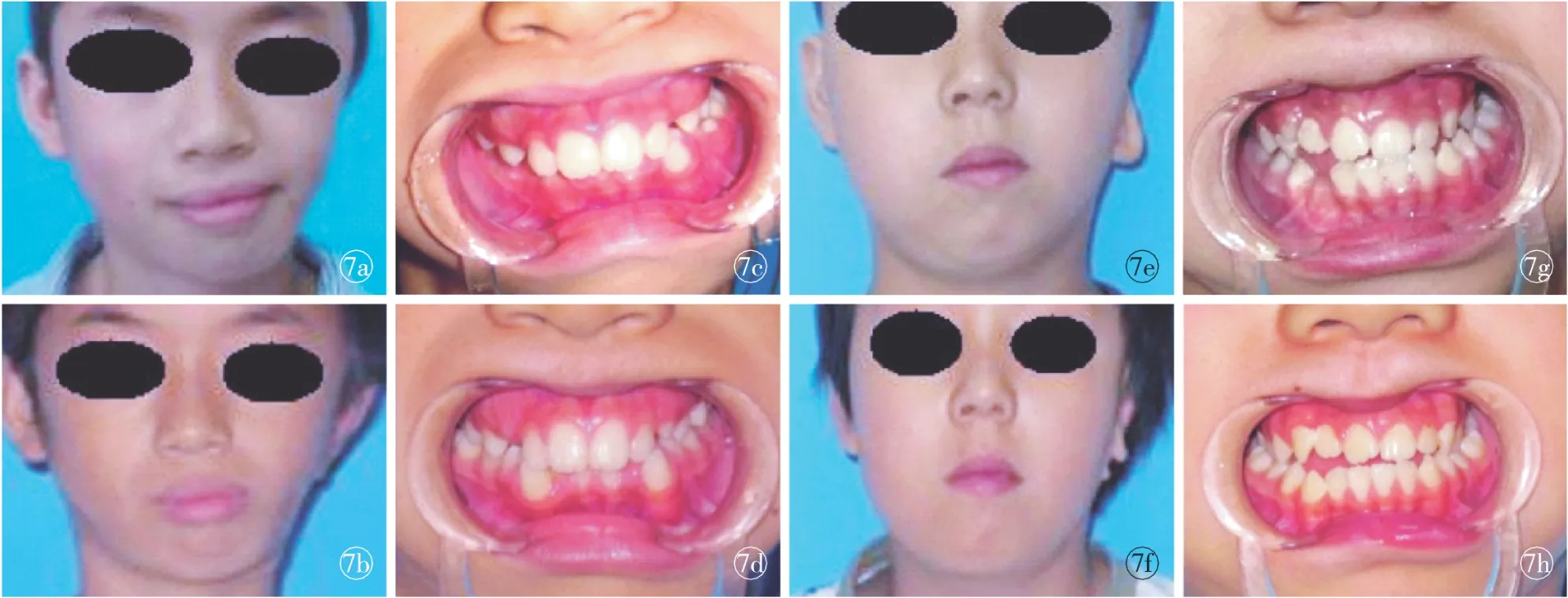
Fig 7 Comparison between preview and postview of child with left hemifacial mircrosomia. a.preview (Case 1). b.postview at 12 months (Case 1). c. preview with occlusion of angle Ⅱ malocclusion (Case 1). d. postview with occlusion at 12 months (Case 1). e. preview (Case 2). f. postview at 6 months (Case 2). g. preview with occlusion of angle Ⅰ occlusion (Case 2). h. postview with occlusion at 6 months (Case 2).
[1] Sakamoto Y, Nakajima H, Ogata H, et al. The use of mandibular body distraction in hemifacial microsomia[J]. Ann Maxillofac Surg, 2013,3(2):178-181.
[2] Dec W, Pehomaki T, Warren SM, et al. The importance of vector selection in preoperative planning of unilateral mandibular distraction[J]. Plast Reconstr Surg, 2008,121(6):2084-2092.
[3] Tuin AJ, Tahiri Y, Paine KM, et al. Clarifying the relationships among the different features of the OMENS+classification in craniofacial microsomia[J]. Plast Reconstr Surg, 2015,135(1):149e-156e.
[4] Ascenço AS, Balbinot P, Junior IM, et al. Mandibular distraction in hemifacial microsomia is not a permanent treatment: a long-term evaluation[J]. J Craniofac Surg, 2014,25(2):352-354.
[5] Sant′Anna EF, Lau GW, Marquezan M, et al. Combined maxillary and mandibular distraction osteogenesis in patients with hemifacial microsomia[J]. Am J Orthod Dentofacial Orthop, 2015,147(5):566 -577.
[6] Tahiri Y, Chang CS, Tuin J, et al. costochondal grafting in craniofacial microsomia[J]. Plast Reconstr Surg, 2015,135(2):530-541.
[7] Meazzini MC, Mazzoleni F, Bozzetti A, et al. Comparison of mandibular vertical growth in hemifacial microsomia patients treated with early distraction or not treated: follow up till the completion of growth[J]. J Craniomaxillofac Surg, 2012,40(2):105-111.
[8] Suh J, Choi TH, Baek SH, et al. Mandibular distraction in unilateral craniofacial microsomia: longitudinal results until the completion of growth[J]. Plast Reconstr Surg, 2013,132(5):1244-1252.
[9] Singh DJ, Glick PH, Bartlett SP. Mandibular deformities: single-vector distraction techniques for a multivector problem[J]. J Craniofac Surg, 2009,20(5):1468-1472.
[10] Shi L, Liu W, Yin L, et al. Surgical guide assistant mandibular distraction osteogenesis and sagittal split osteotomy in the treatment of hemifacial microsomia[J]. J Craniofac Surg, 2015,26(2):498-500.
Discussion on the vector of mandibular distraction osteogenesis for hemifacial mircrosomia
TANGXiao-jun,SHILei,YINLin,etal.
(DepartmentofMaxillofacialSurgery,PlasticSurgeryHospital,ChineseAcademyofMedicalSciencesandPekingUnionMedicalCollege,Beijing100144,China)
Objective To explore the most effective vector of mandibular distraction osteogenesis for hemifacial microsomia using the digital techniques. Methods From 2010 to 2014, 60 patients of hemifacial microsomia were performed the procedures of mandibular distraction osteogenesis using digital techniques. The vector perpendicular to the occlusal plane was applied for the patients with classⅠmalocclusion. The vector should be situated between the occlusal plane and its vertical line for the patients with classⅡ, the angle should be determined using the digital techniques. Results The treatment on 60 cases were successful except for 3 cases with distractor displacement and 5 case with infection. The height of the mandibular ramus and the deviation of the chin were improvement and the cant of occlusal plane was almost corrected. Conclusion The most effective distractive direction is perpendicular to the occlusal plane for patients with angle classⅠ, while, to the patients with angle class Ⅱ, the vector should go between the occlusal plane and its perpendicular line, the precise angle should be made and simulated using the digital techniques.
Hemifcial microsomia; Mandible; Distraction osteogenesis; Digital technique
北京市科委首都临床特色应用研究专项-基于数字化技术的儿童半侧小面畸形早期综合治疗效果评估基金资助项目(Z121107001012112)
作者单位:100144 北京,中国医学科学院北京协和医学院整形外科医院 颌面整形外科中心
唐晓军(1973-),男,黑龙江人,副主任医师,博士.
张智勇,100144,中国医学科学院北京协和医学院整形外科医院 颌面整形外科中心,电子信箱:zhangzh1536@sina.com
10.3969/j.issn.1673-7040.2015.07.004
R782.2
A
1673-7040(2015)07-0394-04
2015-06-10)
猜你喜欢
华西口腔医学杂志(2022年1期)2022-02-14
口腔颌面外科杂志(2021年4期)2021-09-04
四川冶金(2021年6期)2021-02-15
昆明医科大学学报(2021年1期)2021-02-07
科学导报·学术(2020年19期)2020-07-09
锻造与冲压(2020年5期)2020-03-18
中国美容医学(2017年7期)2018-02-02
中国医疗美容(2015年1期)2015-07-12
中国医疗美容(2015年1期)2015-07-12
中国康复(2015年4期)2015-04-10
