A Novel Method for Biological Detection Based on Porous Silicon Multi-Layers Bragg Mirror∗
2018-05-15 00:04MAChengwenLIPengJIAZhenhongLVXiaoyi
新疆大学学报(自然科学版)(中英文) 2018年2期
MA Chengwen,LI Peng,JIA Zhenhong,LV Xiaoyi
(1.School of Physical Science and Technology,Xinjiang University,Urumqi Xinjiang 830046,China;2.School of Information Science and Engineering,Xinjiang University,Urumqi Xinjiang 830046,China)
0 Introduction
Poroussiliconisanattractivebiomaterialbecauseofitseasilymodif i edproperties,suchasporestructure,photonic properties,biocompatibility,biodegradation and surface chemistry.
Porous silicon is the product of electrochemical anodic oxidation of monocrystalline silicon wafers in hydrof l uoric acid electrolyte.By adjusting the etching parameters,aperture control range is about several nanometers to hundreds of nanometers,its inner surface volume ratio,per cubic centimeter can reach hundreds of square meters.It allows a large amount of biological material to be loaded in very small volumes,and it is possible to tailor the pore size and shape as a function of the molecules to be introduced.In addition,the mature biocompatibility and non-toxic PS layer may allow the development of devices directly implanted into organisms without the risk of rejection.Therefore,porous silicon(PSI),as a low-cost,highly sensitive and biocompatible optical biosensor,has attracted much attention and has been studied widely[1−4].
Up to now,porous silicon photonic crystal biosensor has been studied in many aspects.Moreover,a lot of label free optical bio-sensors based on porous silicon materials have been developed[5,6].For example,a biosensor based on grating coupled porous silicon waveguide[7],and using porous silicon waveguide biosensor to detect DNA[8],multilayer bragg mirror[9,10],optical biosensors based on microcavity structure[11,12].In the label free porous silicon biosensor,the porous silicon microcavity structure is more sensitive than that of monolayer and multilayer for biological detection.Lv Xiaoyi uses a porous silicon multi symmetric structure for detecting DNA oligonucleotide biosensors[13].Zhang Hongyan fabricated porous silicon microcavity structure and detected DNA on SOI[14].The above biological detection is based on the shift of the ref l ectance spectrum peak.Li Peng proposed a detection method based on angle spectrum,using porous silicon microcavity structure biosnesor to detect DNA hybridization[15].
In the previous study of porous silicon microcavity biosensors,researchers assume that biomolecules can enter all layers of porous silicon.However,the pore size of porous silicon is about 30∼50nm.For general biomolecules,biomolecules can only enter the f i rst few layers of porous silicon,making it diきcult to enter deeper porous silicon substrates.The sensitivity of porous silicon micro cavity biosensor is limited,which is a problem to be solved for the detection of biological response of porous silicon microcavity devices.Secondly,in order to consider the biological molecules can enter the porous silicon,There are only 6 cycles in the design of the period number of the Bragg mirror.In fact,the more periods number in the design of the Bragg mirror,It has higher sensitivity that the porous silicon photonic crystal biosensor.Based on this,we propose a new method for biological detection based on porous silicon multilayer Bragg mirror.
In this paper,we propose a structure consisting of two diあerent Bragg mirror,the structure has the advantages of combining non destructive with silicon substrate,which ensures the integrity of porous silicon.The integrity of porous silicon is ensured,and then the biological molecules can be directly added into the defect layer,and the defect layer has larger surface pore size,allowing more biological molecules to enter.Real time information of analytes in porous silicon can be measured quantitatively by using optical biological sensing technology,and it has higher sensitivity.
1 Theoretical analysis
The eあective refractive index of the porous silicon layer is def i ned by porosity and refractive index of the pores inside the porous medium.With the increase of the refractive index of the porous space,the eあective refractive index of the PSI layer increases and the optical spectrum of the layer moves toward the long wave direction.Therefore,by monitoring ref l ection or transmission spectra,it is possible to detect the binding of molecules inside the pores,Because the target is captured inside the pore,the refractive index is increased.
In the theoretical calculation,we have designed a porous silicon microcavity consisting of two symmetrical 6 period Bragg mirrors with a total of 25 layers.the entire microcavity structure thickness of about 7.327µm.As shown in Figure 1,the center wavelength of the ref l ection spectrum of the porous silicon microcavity is 1600 nm by the transfer matrix method.When the biomolecule is detected by the porous silicon microcavity,in the ideal state,biological molecules can enter all layers of porous silicon microcavity structures,the average refractive index of each layer increased 0.01,The red shift of the transmission peak wavelength is 11.51nm in the ref l ection spectrum.

Fig 2 theoretical simulation of the angular transmission spectra of biomolecules entering all porous silicon layers
In order to test the potential sensing ability of porous silicon microcavity structure,and to better understand the advantages of PSMC sensor,we use transfer matrix method to calculate the characteristics of PSMC detection in the angle transmission spectrum.As shown in Fig.2,when the incident angle is 19.88 degrees,the transmittance of porous silicon microcavity is the highest in the angular spectrum.By calculating the transfer matrix method,the biomolecules can enter all layers of porous silicon microcavity structure under ideal conditions,the average refractive index of each layer increase 0.01,the incident angle corresponding to the transmission peak of the porous silicon microcavity increases by 2.3 degrees.
In fact,due to the pore size of porous silicon is about 30nm,biomolecules can only enter into the partial layer of porous silicon.With the increase of the layers’of biomolecules penetrating into the porous silicon microcavity,the number of biomolecules can enter the interior is less and less.As shown in Fig.3,when the biomolecules enter the f i rst 6 layers of the microcavity structure,the thickness of the six layers is 1.6665µm,and the refractive index of each layer increase 0.01,and the wavelength of the transmission peak os only res shift 0.94nm by calculating the transfer matrix method.
As shown in Fig.4,when the incident angle is 19.88 degrees,the transmittance of porous silicon microcavity is the highest in the angular spectrum.By the transfer matrix method,when the biomolecules only enter the f i rst 6 layers of the microcavity structure,the thickness of the 6 layers is 1.6665µm,the average refractive index of each layer increase 0.01,the incident angle corresponding to the transmission peak of the porous silicon microcavity increases 0.13 degrees.

Fig 3 theoretical simulation of the change of refl ectance spectrum of biological molecules entering the fi rst 6 layers of porous silicon

Fig 4 theoretical simulation of angular transmission spectra change of biological molecules entering the top 6 layers of porous silicon
Because the biomolecules can only enter the f i rst few layers of the porous silicon microcavity,the microcavity structure can not detect the biomolecules eあectively.Therefore,the traditional PSMC multilayer bragg mirrors has few periods,which makes it easier for entering more biomolecules.But the reduction of the periods number in the multilayer bragg mirror also brings about the following problems.
As shown in Fig.5,the angular transmission spectra of 6 periods and 10 periods porous silicon microcavity structures are calculated by the transfer matrix method.By comparing their FWHM,the less periods number,the half-height width is wider.The quality factor Q of the porous silicon microcavity reduce,which indicates that reducing the number of cycles of the porous silicon microcavity leads to the decrease of the sensitivity of the photonic crystal sensor.
The traditional porous silicon microcavity structure due to biological molecules can only enter the surface layers of porous silicon bragg mirror,and can not enter into all layers of the porous silicon microcavity structure.In order to facilitate the entry of biomolecules,the researchers only fabricated a few number of periods bragg mirrors,this leads to a decrease in sensitivity of biomolecules detection.
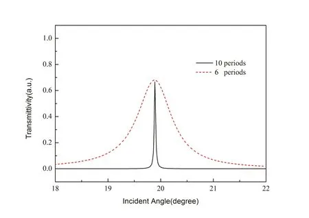
Fig 5 theoretical calculations of angular transmission spectra of porous silicon microcavity structures with 6 and 10 periods

Fig 6 schematic diagram of two separate Bragg mirror combination structures
As shown in Fig.6,based on the above problems.In this paper,a biological detection method based on the structure of porous silicon Bragg ref l ector is proposed,The structure consists of a bragg multilayer and a defect layer,and a symmetrical bragg multilayer.This structure can directly add biomolecules to the defect layer,and there is no limit to the periods’number in the Bragg multilayer.
When the biomolecule only enters the defective layer,assuming that the thickness of the defective layer is 640nm and the refractive index of the defect layer increase 0.01,the wavelength of the transmission peak shifts to 4.29nm by calculating the transfer matrix method.This indicates that the defect layer is still sensitive to the change of refractive index.Although the wavelength shift of the defect in this case is less than 40%of that in the ideal porous silicon all layers,it is only necessary to enter the defect layer with high porosity without entering the bragg multilayers.There is no limit to the layers’number of bragg mirrors.The period number of bragg layer can be farbricated more,which makes the sensitivity of PSMC extends till ten times.
As shown in Figure 7,the porous silicon structure consisting of 10 period Bragg mirrors is calculated by the transfer matrix method.and when the incident angle is within the range of 30 degrees,the maximum refractive index change of the defect layer of porous silicon is 0.2592.

Fig 7 the refractive index change of porous silicon layer with the increase of incident angle

Fig8 Withtheincreaseof theincident angle,the biomolecules enter the refractive index change of diあerent porous silicon layers
As shown in Fig.8,it is shown that by comparing the refractive index changes of the biomolecules entering into the defect layer,the f i rst six layers of the porous silicon,and the all layers of the porous silicon separately.It can be seen that the incident angle is in the range of 1 degree.The dash line in the f i gure indicates that the biological molecules enter the porous silicon under the ideal state,and all layers require the least refractive index change,but this ideal test can not be achieved in the experiment.In the existing porous silicon microcavity sensors,biomolecules can only enter the f i rst 6 layers of porous silicon.By observing the solid line in the f i gure,it can be concluded that the detection sensitivity of the f i rst 6 layers of the porous silicon is the lowest.By observing the dot line in the f i gure,biological molecules enter into the defect layer of porous silicon,compared with the f i rst 6 layers of the porous silicon,the detection of defect layer in porous silicon requires less change of refractive index can achieve the purpose of detection.
2 Conclusion
By theoretical simulation calculations,in the near infrared region,two separate multilayer Bragg ref l ectors are combined.This structure has the advantage of biological molecules can be directly added to the defect layer.Due to the local change of the refractive index of the defect layer,the incident angle shifts,and the interaction of biomolecules can be monitored quantitatively by measuring the amplitude of angular displacement.In the experimental study,it is impossible to realize the total number of biomolecules enter into the porous silicon.In fact,the biomolecules can only enter a few layers,So we propose to add biomolecules directly to the defect layer based on the structure of the Bragg ref l ector,which is more suitable for the current experimental detection studies.The detection method we proposed can be used to detect low concentration biomolecules,and the detection limit can be further improved by the high quality factor Q.
References:
[1]Harraz F A.Porous silicon chemical sensors and biosensors:A review[J].Sensors&Actuators B Chemical,2014,202(10):897-912.
[2]Dhanekar S,Jain S.Porous silicon biosensor:current status.[J].Biosensors&Bioelectronics,2013,41(1):54-64.
[3]Gupta B,Zhu Y,Guan B,et al.Functionalised porous silicon as a biosensor:emphasis on monitoring cells in vivo and in vitro[J].Analyst,2013,138(13):3593-615.
[4]Qiao H,Guan B,Gooding J J,et al.Protease detection using a porous silicon based Bloch surface wave optical biosensor.[J].Optics Express,2010,18(14):15174.
[5]Selena C,Scott R H,Philippe M F,et al.Identif i cation of Gram Negative Bacteria Using Nanoscale Silicon Microcavities[J].Journal of the American Chemical Society,2001,123(47):11797-8.
[6]Chan S,Li Y,Rothberg L J,et al.Nanoscale silicon microcavities for biosensing[J].Materials Science&Engineering C,2001,15(1–2):277-282.
[7]Wei X,Weiss S M.Guided mode biosensor based on grating coupled porous silicon waveguide[J].Optics Express,2011,19(12):11330.
[8]Rong G,Ryckman J D,Mernaugh R L,et al.Label-free porous silicon membrane waveguide for DNA sensing[J].Applied Physics Letters,2008,93(16):161109-1-3.
[9]Huang T H,Pei Y,Zhang D,et al.Patterned porous silicon photonic crystals with modular surface chemistry for spatial control of neural stem cell diあerentiation[J].Nanoscale,2016,8(21):10891-10895.
[10]Volk J,Bal´azs J,T´oth A L,et al.Porous silicon multilayers for sensing by tuneable IR-transmission f i ltering[J].Sensors&Actuators B Chemical,2004,100(1–2):163-167.
[11]Reece P J,Lerondel G,Zheng W H,et al.Optical microcavities with subnanometer linewidths based on porous silicon[J].Applied Physics Letters,2002,81(26):4895-4897.
[12]Ouyang H,Striemer C C,Fauchet P M.Quantitative analysis of the sensitivity of porous silicon optical biosensors[J].Applied Physics Letters,2006,88(16):163108-1-163108-3.
[13]Lv X,Chen L,Zhang H,et al.Hybridization assay of insect antifreezing protein gene by novel multilayered porous silicon nucleic acid biosensor[J].Biosensors&Bioelectronics,2013,39(1):329-333.
[14]Zhang H,Jia Z,Lv X,et al.Porous silicon optical microcavity biosensor on silicon-on-insulator wafer for sensitive DNA detection[J].Biosensors&Bioelectronics,2013,44(1):89-94.
[15]Li P,Jia Z,L¨u G.Hydatid detection using the near-infrared transmission angular spectra of porous silicon microcavity biosensors[J].Sci Reports,2017,7:44798-1-8.