
Arrhythmia and its risk factors post myocardial infarction: A prospective study
2022-02-18 08:10RajinderSharmaIshfaqChowdharyAnkitaSharma
Journal of Acute Disease 2022年1期
Rajinder Sharma, Ishfaq Chowdhary, Ankita Sharma
Department of Medicine, Government Medical College, Jammu, India
ABSTRACT
KEYWORDS: Arrhythmia; Myocardial infarction; Left ventricular ejection fraction
1. Introduction
Cardiovascular diseases (CVDs) (including mainly ischemic heart disease (IHD) as well as stroke) are considered to be one of the main causes of worldwide mortality as well as morbidity, and primary contributors in disability[1]. World Health Organization and Global Burden of Disease study report around 18.6 million deaths from CVD in the past year[2].
Acute myocardial infarction (AMI) and the complications associated with it are considered to be the most life-threatening in atherosclerotic coronary artery diseases (CAD). Most of the mortality in AMI is caused by arrhythmia that include atrioventricular block, bradyarrhythmias, supraventricular tachyarrhythmias, and ventricular arrhythmias[3,4]. Along with arrhythmia present in the acute phase of MI, the risk related to arrhythmia is increased more due to the reopening of an infarctrelated artery; this can lead to serious arrhythmia, eventually increasing the risk of mortality[4]. This has been ascribed to the disturbances in serum electrolytes that are common in the initial 24 h after AMI. Several inorganic salts (particularly alkaline elements) consisting of sodium as well as potassium are implicated among experiments for disbalance in the cardiac rhythm[5].
Though the initial 48-72 h are monitored with caution, less attention is paid to the convalescent period of AMI, whereas patients still are at risk of severe cardiac arrhythmia as well as sudden death[6].
In patients of Indian origin, there is sparse data related to the profile and timing of arrhythmia within 1-7 d of acute MI as well as the factors that increased the probability of such events[7].
Thus the present study was conducted to determine the occurrence of arrhythmia and their associated risk factors in the first week after AMI.
2. Patients and methods
2.1. Study design
A prospective study was conducted in the Department of Medicine of a tertiary care hospital in Jammu over 12 months from November 2018 till October 2019.
2.2. Inclusion and exclusion criteria
All diagnosed cases of AMI were included in the study admitted during the study period. Patients with less than 18 years of age,presence of congenital heart disease, or valvular heart disease were excluded from the study (Figure 1).
2.3. Sample sizes
Sample size calculation based on the study of Shah et al.[8] who observed that the incidence of arrhythmia was 41%. Taking this value as reference, the minimum required sample size with a 10%margin of error and 5% level of significance is 93 patients. To reduce the margin of error, the total sample size taken is 100.
2.4. Ethical consideration
Written informed consent was obtained from all the patients before enrollment and institutional ethical clearance was obtained before beginning the study (IEC/GMC/2019/813, Dated 16.12.2019).
2.5. Diagnosis
The diagnosis of AMI was based on typical signs and symptoms,chest pain, and ECG findings suggestive of ST-segment elevation myocardial infarction (STEMI) for non-STEMI and elevated cardiac biomarkers like creatine phosphokinase and troponin T.
2.6. Data collection
All patients underwent routine blood investigation, 12 lead electrocardiogram, 2D echocardiography, serum electrolytes,cardiac biomarkers, and cardiac monitoring. During the follow-up period of 7 d after AMI, the occurrence of arrhythmia and its types were recorded as an outcome measure. Secondarily, we determined the association of various demographic and clinical parameters with the occurrence of arrhythmia.
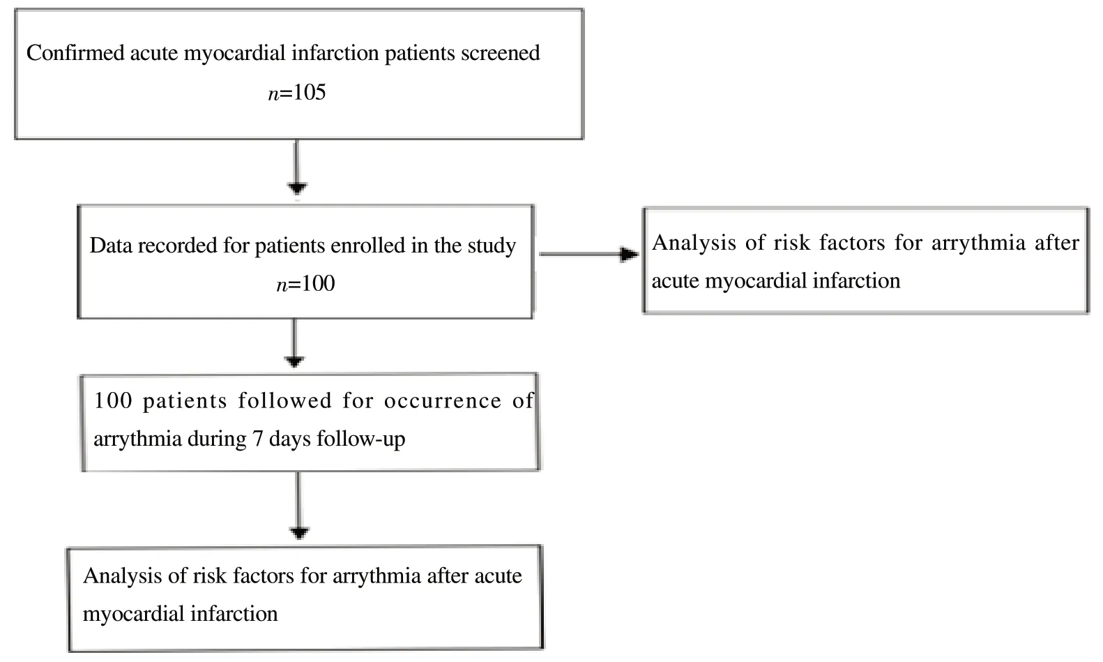
Figure 1. The study flowchart.
2.7. Statistical analysis
The data entry was done in the Microsoft EXCEL spreadsheet and the final analysis was done with the use of Statistical Package for Social Sciences (SPSS) software (IBM manufacturer, Chicago,USA, ver 21.0.). Kolmogorov-Smirnov test was used to determine the normality of the data. The categorical variables was presented in the form of numbers and percentages (%). The quantitative data with normal distribution were presented as the means ± SD. The age data were quantitative and were analyzed using an independent t-test. The comparison of the variables which were qualitative such as gender, ejection fraction, serum sodium, and serum calcium was analyzed using the Chi-square test. If any cell had an expected value of less than 5 then Fisher's exact test was used for the association of serum potassium and magnesium with arrhythmia.Multivariate logistic regression was used to find out significant risk factors of arrhythmia. The significance level of this test was α=0.05.
3. Results
The mean age of the patients was (56.60±12.72) years (range:25-89 years) with the gender distribution of 68% males and 32%females. The comorbidities seen among the study population were hypertension and diabetes. There were 40% smokers who smoked around 8-10 cigarettes per day for a median duration of 5 years,while there were 50% alcoholics who consumed around 90 mL of alcohol daily for a median duration of 2 years. The commonest complaint was chest pain as seen in 94% of cases followed by dyspnea, sweating, vomiting, palpitations, and epigastric pain(Table 1).

Table 1. Demographic and baseline characteristics of the patients.

Table 2. Univariate analysis of parameter associated with arrhythmias .

Table 3. Multivariate logistic regression of significant risk factors of arrhythmia.
Of all 100 cases, 82% had STEMI and 18% had non-STEMI(NSTEMI). It was observed that 34% had extensive anterior wall infarction, 21% had inferior wall infarction, 12% had anteroseptal,19% had inferior wall with right ventricular extension and 6% had anterolateral MI.
Among 100 cases, the incidence of arrhythmia was 73%, and among the 73 cases the most common arrhythmia was sinus tachycardia detected in 30 cases (41.1%), followed by ventricular premature beats in 17 cases (23.2%), sinus bradycardia in 16 cases(21.9%), ventricular tachycardia in 6 cases (5.8%) and atrial fibrillation in 6 cases (8.3%). However, ventricular fibrillation and atrial flutter was not seen in any of the patients.
Of all cases, the incidence of the block was seen in 24 cases out of which 8 had left bundle branch block (33.3%), 6 had right bundle branch block (25.0%), 4 had 2nd-degree heart block (16.7%) and 6 had complete heart block (25.0%).
Most of the arrhythmias were seen between 1-12 h following acute infarction, among which 35.4% of the cases showed arrhythmia immediately within 1 h, 45.6% of cases between 1-12 h, 11.6% between 12 h and 48 h, and 7.4% between 3rd to 5th day.Cardiac failure (ejection fraction<40%) was seen in 51% cases.Ejection fraction, serum calcium, and magnesium were significantly different between non-arrhythmia and arrhythmia patients (P<0.05) (Table 2).
Multivariate logistic regression showed that ejection fraction was an independent significant risk factor of arrhythmia. Patients with ejection fraction >40% had a significantly lower risk of arrhythmia with an adjusted odds ratio of 0.22 (95% CI: 0.08 to 0.64) (Table 3).
4. Discussion
Follow-up of the patients with MI seems necessary because of the occurring of arrhythmia which carry a high morbidity and mortality. In MI non-availability of oxygen causes heart muscles to damage irreversibly. The diastolic and systolic function gets impaired in a MI patient, which increases the susceptibility to arrhythmia. MI results when the heart muscle gets damaged due to a stoppage of blood flow, which increases myocardial metabolic demand, decreases oxygen delivery, decreases delivery of nutrients to the heart's muscles (via coronary circulation)[9].
In our study, there were 27% cases of arrhythmia among which sinus tachycardia was the commonest. This mainly occurs because of ionic alteration and electrolyte disturbances as they disturb the electrical activity of the sinoatrial node causing a block in the conduction, the processes which account for the occurrence of arrhythmia. It must be remembered that arrhythmia is not only a sequel of MI but they are also seen during the process of MI[8].
The mechanisms responsible for cardiac arrhythmia may be divided into disorders of impulse formation, disorders of impulse conduction, or a combination of both[10]. The autonomic nervous system controls the activity of the pacemaker. Systemic factors modulate the action of the pacemaker, which includes endogenous or pharmacological substances and metabolic abnormalities. The parasympathetic system releases acetylcholine, which increases the potassium channel conductivity. It also decreases the activity of myocyte L-type voltage-sensitive calcium channel current, which impacts the rate further down[10].
Failure of conduction of transmitting impulse leads to conduction delay and block. Conduction velocity of an impulse and conduction success is dependent on many factors that include both active and passive membrane properties. These factors are the impulse's stimulating efficacy and the tissue excitability, in which the impulse is conducted[11].
The coupling of the gap junction is a critical factor in determining the safety and velocity of impulse transmission. At high rates,refractoriness is not recovered completely, which leads to impulse blockage. If an impulse reaches a tissue that is still under refractory period, the impulse will not be either conducted or conducted with deviation. This mechanism explains many phenomena like Ashman's phenomenon during atrial fibrillation, block or functional bundle branch conduction of a premature beat, and acceleration-dependent aberration[10].
The occurrence of arrhythmia depends on the wall of the heart that is predominantly affected by MI. Seen in the present study that anterior and the inferior wall MI were the commonest, and the type of arrhythmia commonly seen were sinus tachycardia, ventricular premature beats, and sinus bradycardia with various types of conduction blocks. The findings were in line with various other studies where anterior and inferior wall MI was the commonest,and sinus tachycardia with ventricular premature beats was significantly associated with it[8].
In addition, the left ventricular ejection fraction after MI becomes an important predictor of arrhythmia. We determined that if the left ventricular ejection fraction was more than 40% there was significantly less chance of the occurrence of arrhythmia. This indirectly shows that the heart muscles are working in good conditions and the electrical conductivity has been maintained.Besides one may also use global longitudinal strain as a marker to assess the recovery of cardiac muscles while predicting the occurrence of arrhythmia however future studies are recommended to validate that same.
Besides, electrolyte levels also have a significant impact on the prognosis of patients with myocardial infarction. AMI has been reported to be monitored using changes in electrolyte levels. There are various types of electrolytes present in the body, each with a distinct and significant function; however, majority are involved in the maintenance of the fluid balance between the intracellular(within the cell) and extracellular (outside the cell) environments[9].
It is important to maintain balance as it is helpful in the maintenance of hydration, nerve impulses, normal functioning of muscles, and maintaining pH level. The main electrolytes present in the body include sodium, potassium, magnesium, calcium,and chloride. The significant factors for the determination of electrophysiological properties related to the myocardial membrane are serum sodium, potassium, and chloride[9].
Among the various electrolyte disturbances, hypocalcemia and hypomagnesemia showed a significantly higher risk in association with arrhythmia. Calcium maintains depolarization and is involved in myocardial contractility while magnesium stabilizes the cell membrane and acts in concert with potassium and is a calcium antagonist. It dilates coronary arteries, peripheral systemic arteries and reduces afterload. Few studies have been done till now and less information is available in the literature about the prognostic value of serum electrolytes in ischemic heart diseases. Patil et al.[12]reported that in patients with AMI, maximum electrolyte imbalance was present in calcium. In about 50% of cases, hypocalcemia was noted and one of the studies[13] showed a correlation between hypomagnesemia and ventricular arrhythmia.
Though not seen in our study, sodium and potassium (two of the complex electrolytes present in the body) have also been seen to be associated with arrhythmia[14-16]. Wali et al.[16] in a case-control study (n=50), including patients with AMI, found a significant reduction in levels of sodium and potassium among cases. Similar findings were reported by Hariprasad et al.[17] as it was found that AMI patients had reduced levels of sodium and potassium.Some studies partly corroborated with the study in finding no association of sodium or potassium levels with the occurrence of arrhythmia[17].
A study by Verma et al.[5], including 75 patients with AMI with or without arrhythmia, found that concentration of serum sodium was unaffected among patients having AMI with or without arrhythmia while the concentration of serum potassium was significantly reduced among patients with AMI with arrhythmia.
Ventricular arrhythmia as well as consequent sudden cardiac death because of the AMI are the most frequent causes of death among humans. Lethal ventricular arrhythmia, such as ventricular fibrillation (VF), before hospitalization is reported to be present in >10% of all the cases of AMI with the survival among such patients being poor[18].
Therefore, recognizing the risk factors, as well as mechanisms related to VF after AMI is significant, which can aid in the implementation of novel risk stratification models and therapeutic methods for decreasing mortality among individuals having high CV risk. Usually, evaluation of spontaneous VF after AMI is difficult because it generally takes place unexpectedly among the low-risk subgroup[18]. However, our results show disturbed electrolyte disturbances in the initial 7 d following AMI which may provoke arrhythmia and increase morbidity and mortality.
Though there was no mortality observed in the present study but it still amounts to a limitation. The treatment protocol was not recorded in the present study. Lastly, the study population sample size was small.
It can be concluded that arrhythmia is a common occurrence in the initial follow-up week after myocardial infarction. The type of arrhythmia and the type of block may depend on the heart muscles involved during myocardial infarction. Ejection fraction is a significant risk factor that may affect the occurrence of arrhythmia.
Conflict of interest statement
The authors report no conflict of interest.
Authors'contributions
R.S.: Concept, design, literature search, data analysis, manuscript preparation; I.C.: Design, Data acquisition, manuscript review;A.S.: Design, Data acquisition, manuscript review.
 Journal of Acute Disease2022年1期
- Journal of Acute Disease的其它文章
- Incidence of adverse reactions to COVID-19 vaccination: A metaanalysis of randomized controlled trials
- Health literacy, behavioral and psychosocial characteristics in coronary artery patients: A hospital-based study in Turkey
- Acute and sub-acute toxicities of hydroalcoholic extract of Allium affine aerial parts in rats