
Reduced choroidal peripapillary capillaries in thyroidassociated ophthalmopathy with early stage of dysthyroid optic neuropathy
2022-07-30 10:02JiaHuiWuLiYingLuoHaoZhouYingWuJianZhangJinWeiCheng
INTRODUCTION
Thyroid-associated ophthalmopathy (TAO), also known as Graves’ ophthalmopathy, is an autoimmune disorder which has characteristic ocular manifestations,such as proptosis, eyelid retraction, eyelid lag and restrictive extraocular myopathy
. Among a series of TAO associated clinical findings, dysthyroid optic neuropathy (DON) is the most severe vision-threatening condition. The pathogenesis of DON is complicated, which is not fully understood by far.The most widely accepted theory is that DON is caused by the combined effects of mechanical, vascular and inflammatory process. Mostly, DON is secondary to a compartment syndrome in orbital apex, which is caused by enlargement of extraocular muscles and orbital fat resulting from orbital fibroblast deposition of hyaluronic acid
.
Our study demonstrated that a significant reduction of choroidal RPC observed in DON patients rather than normal and TAO without DON by using OCTA. RPC is a superficial capillary layer which comprise a unique vascular plexus. There have been reports demonstrating that RPC is necessary to metabolic demands of retinal ganglion cell (RGC) axons
.As the RGC axons are vulnerable to decease of blood flow, the structural changes to RPC network could lead to pathogenesis of RGC axonal loss
. There is evidence also showing an correlation between RPC loss and RNFL changes in chronic glaucoma
. The change of peripapillary microvasculature could not be efficiently detected by OCT
, which indicates that OCTA could be the most effective and easiest way to detect changes in retinal and/or choroidal microvasculature.Our data indicated a significant correlation between RPC reduction and VF defect. However, the decrease of choroidal RPC density could not be reversed 6mo after relieving optic nerve compression by either corticosteroid treatment or optic nerve decompression surgery though vision acuity significantly improved. However, longer term follow-up is required to further understand if the reduction of choroidal RPC could be recovered or permanently affected. Therefore, it is suggesting that choroidal RPC density could be clinically useful for early diagnose of DON.
There are around 4% to 8% TAO patients having DON
,of which the irreversible vision loss is largely caused by delayed diagnosis due to lack of efficient detecting method at early stage of TAO. It has been reported that the axonal changes of optic nerve are detected in patients with DON
.The compression caused by enlarged extraocular muscles and orbital fat may stretch the optic nerve and reduce the blood flow supply of retina. A report demonstrated that blood flow volume of superior ophthalmic vein decreased in DON eyes
.These findings suggest that the changes of optic nerve and hemodynamic state of the eye might be valuable for early diagnosis of DON.
There is no specific diagnosis guideline for DON due to various of clinical manifestations and limited detecting methods. Most clinicians diagnose DON by a combination of radiological findings and clinical manifestations. However, some patients may not present external signs, such as proptosis, because in some cases, DON only caused by congestion at the orbital apex
. The decline of visual acuity also sometimes lag behind other clinical presentations of DON, therefore, a number of tests should be run to evaluate the function of optic nerve for diagnosis of DON, including papillary exam, automated visual field (VF) and contrast sensitivity
.
图画书讲读,应该建立在对图画书艺术的理解和把握的基础上。儿童对图画书的喜爱,与图画书的内容、形式、表达,与其图画文字的共同讲述故事,与其独有的艺术设计和效果,都有着密不可分的关系。图画书因此建立起不同于其他读物的欣赏方式,儿童更有自己进入图画书艺术世界的方法和途径,有符合他们心理和趣味的审美体验。教师进行图画书的讲读活动,应尽可能在认识图画书艺术构成和特点的基础上进行,帮助儿童积累或调动他们自己的阅读经验,尽可能充分地、全方位地欣赏图画书作品,获得愉悦并提升审美能力。
Optical coherence tomography angiography (OCTA) is a noninvasive imaging facility which could be applied to measure the thickness of retinal nerve fiber layer (RNFL), macular and many other parameters of the eye. OCTA is a high-speed optical coherence tomography (OCT) that could characterize a map of blood flow and vessel network of different layer and area of the retina and choroid, which is achieved by comparing the captured signals between sequential scans taken at same crosssection. OCTA assists clinicians to non-invasively visualize and assess retinal and choroidal perfusion, while at the same time the assessment is trustworthy due to its reliable reproducibility,sensitivity and specificity
. With the emergence of OCTA,it is possible to study the correlation between microvascular perfusion changes and the development of DON.
Here we demonstrated that OCTA could detect early defects in density of radial peripapillary capillaries (RPC) around optic disc in DON, which was also significantly correlated with defect of VF and vision. The findings suggested that OCTA could be used to early diagnose DON in clinics.
SUBJECTS AND METHODS
Ethical Approval This was a retrospective cross-sectional study conducted at the Department of Ophthalmology in Shanghai General Hospital, China, from January 2019 to December 2020. The study protocol and ethics were approved by the Ethics Committee of Shanghai General Hospital (ref 2020KY206). The study was conducted complied with the tenets of the Declaration of Helsinki. Written informed consent was obtained from all participants.
李闺女又呸了一口:李六如,真是越来越不知道羞耻了。原先拆迁时,你想当个先进也就算了。如今,又勾结佟金鑫占了那点口粮田。你说,老少爷们今后怎么活?
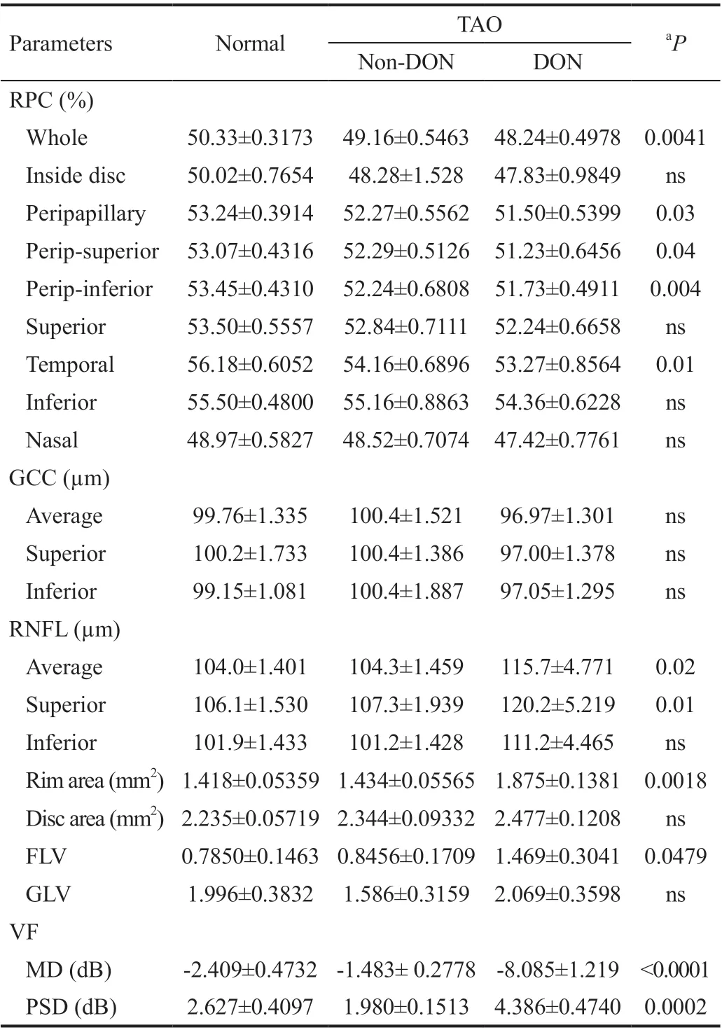
Significant Reduction of Radial Peripapillary Capillaries in Dysthroid Optic Neuropathy Next, we further divided the patients of TAO into two subgroups, DON and non-DON,according to the criteria described in method section. DON had a significant reduction in both VF and the percentage of choroidal RPC, including whole and peripapillary, compared to both normal and TAO without DON (Figure 1). The percentage of whole choroidal RPC significantly reduced from 50.33%±0.3173% in normal to 49.16%±0.5463% in TAO without DON and further to 48.24%±0.4978% in DON(
=0.0041). The percentage of peripapillary choroidal RPC was also significant reduced from 53.24%±0.3914% in normal to 52.27%±0.5562% in TAO without DON, and further reduced to 51.50%±0.5399% in DON (
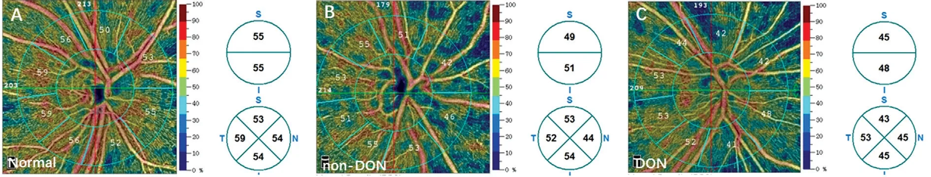
=0.03; Table 3). In agreement to the findings, the representative OCTA images also illustrated that the choroidal capillaries around optic disc were thinner in DON patients than that in normal and TAO without DON (Figure 2). There was no significant difference between DON, TAO without DON and normal controls in most tested parameters (Table 3). The above data suggested that change of choroidal RPC could be a specific sign in DON,and there was a possibility that the reduction of choroidal RPC in DON correlates with VF defect.
Patients diagnosed with TAO were based on Barley criteria
.Inclusion criteria for TAO participant were: 1) At least 18 years of age; 2) No history of radioactive iodine therapy or thyroidectomy. TAO participants were further divided into two groups, DON and non-DON. The diagnosis of DON was based on clinical findings
: 1) Decreased visual acuity compared to previous medical records; 2) Apparent VF defect mean deviation (MD) <-10 dB in Humphrey test; 3) Relative afferent pupillary defect; 4) Evidence of apical crowding in computed tomography or magnetic resonance imaging.
RTVue-XR Avanti system has been designed to minimize scanning time based on the SSADA algorithm. The system can also measure the variation of OCT signals among consecutive scans, therefore the motion of blood flow could be captured. In order to quantify and analyze the nerve fiber layer and macular circulation,
retinal angiogram images were processed and vessel density was calculated using the Avanti trend analysis software.
Exclusion criteria for all participants were: 1) Any retinal pathology and optic neuropathy, such as uveitis and diabetic retinopathy; 2) Any complication inducing VF loss, such as glaucoma or ocular tumor; 3) Any history of ocular trauma or intraocular surgery; 4) Vulnerable individuals or those who cannot conduct any test required in this study.
Humphrey Visual Field Test Humphrey Visual Field Analyzer II 750 (Carl Zeiss Meditec) was used to test VF for all participants, the data was calculated by Humphrey Swedish Interactive Threshold Algorithm (SITA) 30-2 test.The included results should meet the criteria that fixation loss was less than 20%, and both false-negative errors and falsepositive errors were less than 15%. All tests were performed without any inappropriate operation, such as eyelid artefacts,inattention, and fatigue effects. Any defects of VF caused by other diseases was excluded as described before.
通过本次研究可以看出,凶险性前置胎盘患者相对于普通前置胎盘患者来说,面临的威胁更大,可能导致的不良妊娠结局与不良新生儿结局的概率更高。另外,在一般资料对比中我们可以看出,高龄产妇、孕次和产次较高的产妇其发生凶险型前置胎盘的可能性更大,因此,临床上对于此类前置胎盘产妇要给予特别关注。
OCTA Image Acquisition and Processing The RTVue-XR Avanti could be used to visualize vascular structures of distinct layers of the retina and choroid. The scan was non-invasive and achieved by low-coherence interferometry. RTVue-XR Avanti used an 840 nm light source as the scan beam wavelength with an A-scan rate of 70 000 A-scan per second. Motion artefacts were minimized by dual orthogonal volumetric imaging of the retina. Each studied eye received four volumetric raster scans,which were composed of two horizontal priority (X-fast)and two vertical priority (Y-fast) scans. The scans provided consecutively information outside a 6×6 mm
field size of observed structures and assessed the details of retina in distinct layers.
Inclusion criteria for normal subjects were: 1) at least 18 years of age; 2) normal clinical appearance of the optic disc; 3) no RNFL loss; 4) no VF defect.
Measurement of Radial Peripapillary Capillaries, Macular Ganglion Cell Complex and Peripapillary RNFL The thickness of peripapillary RNFL and macular GCC were obtained using RNFL 4.5 scanning mode and analyzed by the GCC scan algorithm installed in the RTVue-XR Avanti. The RNFL thickness was measured around the optic disc with a circle of 4.5 mm diameter using RNFL mode, while the GCC scan was examined around the fovea with a square area of 6×6 mm
.GCC scan covers multiple layers of the retina, including the RNFL, the ganglion cell layer and the inner plexiform layer.The calculation of the scanned segmentations of the RNFL and GCC was analyzed by the SSADA algorithm. Furthermore,the SSADA algorithm can also measure and analyze separated areas of the retina including average, superior and inferior hemi-retinal RNFL and GCC.
Statistical Analysis All the data were calculated as mean and standard deviations and compared between groups. For the comparison between two groups, Student’s
test was used to compare the average values of all the measurements. Gender frequency comparison was analyzed by the Chi-square test.The correlation analysis was performed by univariate analysis with Pearson correlation test and multivariate analysis with ANOVA testing to determine the correlation between RPC and other parameters, such as visual acuity and MD and pattern standard deviation (PSD) of VF. Statistical significance was considered as
<0.05. One-way ANOVA test with Tukey correction was applied for multiple comparison among groups with resultant significance level set at
<0.01.
RESULTS
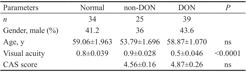
Demographic Data and Clinical Features Table 1 summarizes the clinical characteristics of each group. According to the inclusive and exclusive criteria, 34 eyes were included in normal group, while 64 eyes were included in TAO group.TAO was further divided into two groups, DON and TAO without DON, with 39 eyes in DON and 25 eyes in non-DON respectively. There was no statistically significant difference in DON, TAO without DON and normal controls, regarding to age and gender. The visual acuity of DON was significantlylower than the other two groups, though the CAS showed no difference between non-DON and DON.
据世界卫生组织首份《全球糖尿病报告》显示,全球糖尿病患者人数从1980年的1.08亿增加到2014年的4.22亿。目前我国确诊的糖尿病患者已经超过1亿,居世界首位。据统计,糖尿病在我国的发病率已经超过11.2%,处于糖尿病前期的人数占总人口的50.1%,我国约70%的糖尿病患者不知道自己已经患上糖尿病。而接受治疗的成人糖尿病患者中,血糖控制率不到40%。许多糖尿病患者服用了几十年的降糖药也没有把血糖降下来,药越吃越多,血糖却越来越高,各种并发症越来越严重。
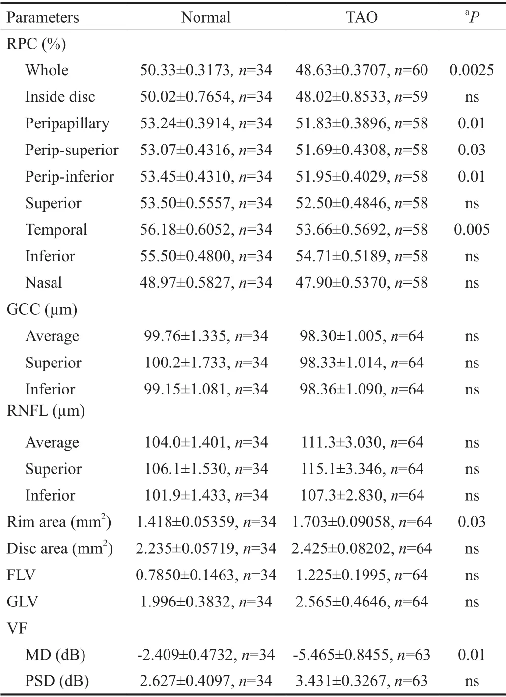
Reduction of Radial Peripapillary Capillaries in Thyroid Associated Ophthalmopathy Compared to Normal Table 2 is an overview of a comparison in a series of parameters between TAO and normal subjects. When compared to normal participants, TAO showed significant reduction in the percentage of choroidal RPC, including whole and peripapillary area of choroidal RPC, as well as in MD of VF (Table 2). The percentage of whole choroidal RPC was 50.33%±0.3173%in normal controls, but reduced to 48.63%±0.3707% in TAO(
=0.0025). Similarly, significant reduction was seen in peripapillary choroidal RPC (51.83%±0.3896% in TAO
53.24%±0.3914% in normal controls,
=0.01; Table 2). While there was no statistical difference between the two groups in thickness of GCC and RNFL, disc area and percentage of focal loss volume (FLV) and global loss volume (GLV).

Study Participants A total number of 98 eyes from 50 subjects were enrolled according to the criteria of this study and imaged by RTVue-XR Avanti (Opto Vue, Inc, Fremnt, CA,USA) platform and analyzed with the split-spectrum amplitude decorrelation angiography (SSADA) algorithm. Each patient underwent a series of ophthalmological examinations, including slit-lamp clinical examination, best-corrected visual acuity test, ocular motility, severity of proptosis, VF test, clinical activity score (CAS)
and OCTA scanning of peripapillary RNFL, macular ganglion cell complex (GCC) and choroid RPC. There were 11 DON patients received intravenous corticosteroids treatment to relieve the clinical symptoms of optic nerve compression, while 5 of whom further received orbital decompression surgery due to the unresponsive to corticosteroids treatment. For the eyes received steroid therapy and surgery, the examinations were practiced both pre- and post- treatment/surgery. The comparison was made between pre-operation and 6-month after treatment/surgery.
Orbital imaging techniques, such as MRI and CT, play a vital role in diagnosing and following DON
. The muscle index is significantly greater in orbits with DON, and DON almost never occurs in patients when muscle index is less than 50%
.Orbital soft tissue imaging also help to diagnose DON, with up to 94% sensitivity and 91% specificity
. However, cost of these imaging detecting methods is high and some disable patients may find it difficult to take the scans. Therefore, there is still a demanding of a new technique which could detect subtle changes for early diagnose of DON.
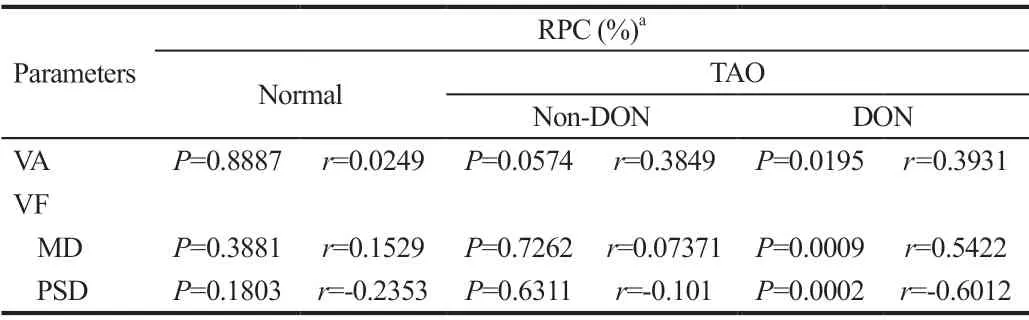
Correlations Between Radial Peripapillary Capillaries and Visual Function Parameters To further investigate if the reduction of choroidal RPC accounts for any pathological changes in DON, we ran a series analysis to detect any correlations. Data showed that the whole percentage of choroidal RPC had a significant correlation with visual defect,especially defect in VF in DON (
=0.5422), while such correlations were not observed in either normal (
=0.1529) or TAO without DON (
=0.07371; Table 4), which suggested that the reduction of choroidal RPC was a specific change in DON.Radial Peripapillary Capillaries Density Could not be Reversed by Medical or Surgical Decompression To investigate if the RPC density and other parameters would improve when DON is relieved, we compare all these observed clinical data before and after treatment. There were 11 DON eyes received intravenous methylprednisolone pulse therapy due to severe symptoms of optic nerve compression such as rapidly decline of vision acuity and defect of VF, but 5 of which did not respond well to corticosteroids treatment as the deterioration of vision and VF continued. Therefore,the 5 DON eyes further received orbital decompression surgery followed with symptoms of optic nerve compression significantly improved in all cases. After medical or surgical decompression, vision acuity and VF improved in all treated eyes, but there was no significant difference of choroidal RPCdensity between pre-treatment (whole 48.28%±1.147%) and post-treatment (whole 47.58%±1.144%; Table 5). The data demonstrated that both medical and surgical decompression could reverse defected visual function to a certain level, but the decline of choroidal RPC still remained.
DISCUSSION
In this study, we demonstrated that the percentage of choroidal RPC was significantly reduced in the patients of DON compared to both normal controls and the patients of TAO without DON. The reduction of choroidal RPC also correlated with defects of VF, which suggested the change of choroidal RPC could be an early sign of optic nerve damage. OCTA can be used to investigate vessel volume and density of multiple layers of the eye, which makes clinicians able to observe changes undetected by other investigating methods.
So far, there is no single protocol or guideline for diagnosing DON
, clinicians have to measure grades of clinicalsigns and symptoms of patients, and also run a series of tests, including CT or MRI scan, and VF test. Only with the combination of clinical findings and test results can DON be diagnosed. However, there are still a number of DON patients facing with delayed diagnosis due to unspecific clinical presentation
. For example, some patients with DON only caused by congestion at orbital apex without any sign of proptosis, and patients sometimes are lack of clinical manifestations of orbital inflammation, which could cause the delay use of radiology test as there is little sign of further investigations. Decreased visual acuity is also an unspecific symptom, though it is more often found in DON rather than thyroid eye diseases (TED) alone. It has been reported that there are about 47% of DON patients with visual acuity lower than 20/40, while the number is only 3% in TED patients
.Reduction of contrast sensitivity, colour vison change and an afferent pupillary defect are the other signs specific for DON,though all of these signs can be absent in some cases
.

VF test can accurately detect DON, according to studies, most of the DON eyes develop a central or paracentral scotoma during the progress of disease
. Visual evoked potential(VEP) can also assist to detect DON, and sometimes it is even more sensitive than VF test
. Therefore, the retinal function is one of the diagnostic factor for DON. Our data also suggested that there was a significant difference of VF defect between DON and non-DON.
大老李已经五十多岁了,还从五十里外的西平老家来这里下井。他的背微驼,和我们常常见到的那种老实巴交的高个子男人一样,整天不怎么言语。他的大女儿读高中,儿子小学。大老李极疼他的孩子,他常常盯着一张皱巴巴的照片看,那是他们的全家福。他还买些纸笔书包的存着,说等到回家的时候给孩子带回去。他说,现在农村的孩子们除了读书这条道再没其他出路了。看来他是极想把自己的孩子培养成大学生的。然而此刻,西山轰隆一声,一缕灰烟过后,大老李两腿一蹬,双眼一闭,唉,都他妈的过去了。完蛋了,一切都完蛋了。
(一)释义的内容要能够清楚地标注出词义中是借代意义的义项,而不应该把借代意义混同为词的普通一般的指称意义。比如:
There were studies indicating that the macular microvascular densities were significantly reduced in TAO patients
. But our data presented no significant finding observed neither between normal and TAO, nor between normal and DON.One of the possible reasons of getting different result is that the acquisition area of GCC is different, we acquired 6×6 mm
OCTA images for GCC rather than 3×3 mm
acquired in other studies. Another possibility is that the different enrolled criteria of TAO may give biased readout. There was even one study reporting that microvascular density significantly increased in active TAO
. Taken these findings together, the GCC density varies during different stage of TAO, which indicates that the change of GCC density could not effectively monitor development of TAO.
Our data demonstrated that the density of choroidal RPC,both whole and peripapillary area, was significantly decreased in DON compared to normal eyes and TAO without DON.The decrease of vessel density in the peripapillary area in eye with DON had also been shown by one previous study
. The reduction of choroidal RPC has been found to be correlated with defects of VF. Therefore, the change of choroidal RPC could be an early sign of optic nerve damage. Furthermore,our study found that the reduction of RPC density could not be reversed by medical or surgical decompression. So far, the most widely accepted mechanism is that DON is secondary to a compartment syndrome in orbital apex, and the changes of vessels in orbit may also be related to DON. The decreased blood flow in the active stage of orbitopathy, while the reversed or even absent blood flow in many advanced cases can even induce the optic nerve vasculature which further develops ischemia. Medical and surgical decompression could decrease the direct optic nerve compression in DON, however,might not benefit to restore the vasculature
.
还记得那是去年十月的最后一天中午,我们准时来到了“神秘”的校长室。校长室里宽敞明亮,一尘不染,陈校长已经在那里微笑地看着我们。在听完我们简单的自我介绍后,陈校长笑着说:“我非常高兴能接受你们的采访,你们有什么问题吗?”望着陈校长那慈祥的笑容,我们紧张的情绪一扫而空,紧接着就开始了关于校庆三十周年的专访。
疫情对生猪生产产生了一定影响,春节前猪价或出现一波上涨。主要是由于部分地区仔猪无法调运。目前很多自繁自养和公司+农户企业生猪育肥需要跨市、跨省调运仔猪,由于仔猪无法调运,特别是9月中旬之后,导致繁育场压栏严重、仔猪死亡率较高,而育肥场无猪可育,因此将会影响4个月以后的生猪供应,或将导致某个时期猪价较快上涨,2019年二季度猪价淡季不淡。
There are limitations in our study. First, the observation period is not very long. Longer follow-up could help to get more comprehensive understanding of the role of choroidal RPC in the development of DON. Second, the sample size of treated eyes is also small, more samples would make the conclusion stronger. Last, the diameter of analyzed area by OCTA seems to make some biases in making conclusions, which requires further investigations in future study.
In conclusion, there are advantages of using OCTA in diagnosis of DON. It improves our understanding of pathogenic relationships between optic disc circulation and VF defect. It is also non-invasive and easy to operate, which can be accepted by a wide range of patients. Therefore, choroidal RPC scan by OCTA could be an effective way to investigate and monitor DON, it is also possible to help in early diagnosis of DON.
ACKNOWLEDGEMENTS
Authors’ contributions: Cheng JW designed the study,wrote the manuscript and collected data; Wu JH wrote the manuscript, collected and analyzed data; Luo LY, Zhou H, Wu Y, Zhang J, Cheng JW performed OCTA and collected data.All authors have read and approved the manuscript.
Fe0-PRB技术在含铀废水处理方面得到了研究与应用,但也存在许多缺陷与不足,制约了该技术的进一步发展和实际应用的推广.因此,对这些问题的研究与改进,将会成为Fe0-PRB技术在含铀废水处理方面的研发重点.
Supported by the National Natural Science Foundation of China (No.81170874; No.81900868).
Conflicts of Interest: Wu JH, None; Luo LY, None; Zhou H,None; Wu Y, None; Zhang J, None; Cheng JW, None.
1 Bartley GB, Gorman CA. Diagnostic criteria for Graves’ophthalmopathy.
1995;119(6):792-795.
2 Neigel JM, Rootman J, Belkin RI, Nugent RA, Drance SM, Beattie CW,Spinelli JA. Dysthyroid optic neuropathy. The crowded orbital apex syndrome.
1988;95(11):1515-1521.
3 Victores AJ, Takashima M. Thyroid eye disease: optic neuropathy and orbital decompression.
2016;56(1):69-79.
4 Blandford AD, Zhang D, Chundury RV, Perry JD. Dysthyroid optic neuropathy: update on pathogenesis, diagnosis, and management.
2017;12(2):111-121.
5 Zhang T, Xiao W, Ye HJ, Chen RX, Mao YX, Yang HS. Peripapillary and macular vessel density in dysthyroid optic neuropathy: an optical coherence tomography angiography study.
2019;60(6):1863-1869.
6 Özkan B, Anik Y, Katre B, Altintaş Ö, Gençtürk M, Yüksel N.Quantitative assessment of optic nerve with diffusion tensor imaging in patients with thyroid orbitopathy.
2015;31(5):391-395.
7 Konuk O, Onaran Z, Ozhan Oktar S, Yucel C, Unal M. Intraocular pressure and superior ophthalmic vein blood flow velocity in Graves’orbitopathy: relation with the clinical features.
2009;247(11):1555-1559.
8 Jia YL, Wei E, Wang XG, Zhang XB, Morrison JC, Parikh M, Lombardi LH, Gattey DM, Armour RL, Edmunds B, Kraus MF, Fujimoto JG,Huang D. Optical coherence tomography angiography of optic disc perfusion in glaucoma.
2014;121(7):1322-1332.
9 Wang X, Jiang C, Ko T, Kong X, Yu X, Min W, Shi G, Sun X.Correlation between optic disc perfusion and glaucomatous severity in patients with open-angle glaucoma: an optical coherence tomography angiography study.
2015;253(9):1557-1564.
10 McKeag D, Lane C, Lazarus JH,
; European Group on Graves’Orbitopathy (EUGOGO). Clinical features of dysthyroid optic neuropathy: a European Group on Graves’ Orbitopathy (EUGOGO)survey.
2007;91(4):455-458.
11 Bartalena L, Baldeschi L, Dickinson AJ,
Consensus statement of the European group on Graves’ orbitopathy (EUGOGO) on management of Graves’ orbitopathy.
2008;18(3):333-346.
12 Dolman PJ, Rootman J. VISA classification for Graves orbitopathy.
2006;22(5):319-324.
13 Beden Ü, Kaya S, Yeter V, Erkan D. Contrast sensitivity of thyroid associated ophthalmopathy patients without obvious optic neuropathy.
2013;2013:943789.
14 Jeon C, Shin JH, Woo KI, Kim YD. Clinical profile and visual outcomes after treatment in patients with dysthyroid optic neuropathy.
2012;26(2):73-79.
15 Lipski A, Eckstein A, Esser J, Loesch C, Mann K, Mohr C, Jurklies B.Course of pattern-reversed visual evoked cortical potentials in 30 eyes after bony orbital decompression in dysthyroid optic neuropathy.
2011;95(2):222-226.
16 Barrett L, Glatt HJ, Burde RM, Gado MH. Optic nerve dysfunction in thyroid eye disease: CT.
1988;167(2):503-507.
17 Lima B, Perry JD. Superior ophthalmic vein enlargement and increased muscle index in dysthyroid optic neuropathy.
2013;29(3):147-149.
18 Giaconi JA, Kazim M, Rho T, Pfaff C. CT scan evidence of dysthyroid optic neuropathy.
2002;18(3):177-182.
19 Yu PK, Cringle SJ, Yu DY. Correlation between the radial peripapillary capillaries and the retinal nerve fibre layer in the normal human retina.
2014;129:83-92.
20 Scoles D, Gray DC, Hunter JJ, Wolfe R, Gee BP, Geng Y, Masella BD,Libby RT, Russell S, Williams DR, Merigan WH.
-
imaging of retinal nerve fiber layer vasculature: imaging histology comparison.
2009;9:9.
21 Mammo Z, Heisler M, Balaratnasingam C,
. Quantitative optical coherence tomography angiography of radial peripapillary capillaries in glaucoma, glaucoma suspect, and normal eyes.
2016;170:41-49.
22 Fard MA, Suwan, Moghimi S, Geyman LS, Chui TY, Rosen RB, Ritch R. Pattern of peripapillary capillary density loss in ischemic optic neuropathy compared to that in primary open-angle glaucoma.
2018;13(1):e0189237.
23 Chien JL, Sioufi K, Ferenczy SR, Say EAT, Shields CL. Optical coherence tomography angiography detects subclinical radial peripapillary capillary density reduction after plaque radiotherapy for choroidal melanoma.
2020;40(9):1774-1782.
24 Wu YF, Tu YH, Bao LL, Wu CM, Zheng JW, Wang JH, Lu F, Shen MX, Chen Q. Reduced retinal microvascular density related to activity status and serum antibodies in patients with Graves’ ophthalmopathy.
2020;45(5):576-584.
25 Ye L, Zhou SS, Yang WL, Bao J, Jiang N, Min YL, Yuan Q, Tan G,Shen M, Shao Y. Retinal microvasculature alteration in active thyroidassociated ophthalmopathy.
2018;24(7):658-667.
猜你喜欢
今日农业(2022年14期)2022-09-15
农村百事通(2022年4期)2022-04-22
作文·初中版(2021年8期)2021-09-13
爱你·健康读本(2019年2期)2019-06-11
中国绿色画报(2017年8期)2017-09-01
湖北畜牧兽医(2016年8期)2016-11-21
农家顾问(2016年6期)2016-05-14
中国动物保健(2015年4期)2015-10-21
集装箱化(2014年11期)2014-12-17
江苏教育(2009年22期)2009-07-20
 International Journal of Ophthalmology2022年7期
- International Journal of Ophthalmology的其它文章
- Impact of OCT scan-patterns in identifying morphological features of lamellar macular holes and macular pseudoholes
- Virtual reality training improves accommodative facility and accommodative range
- Short-term effect of 0.01% atropine sulphate eye gel on myopia progression in children
- Incidence of ocular manifestations in patients with graft versus host disease after allogeneic stem cell transplant in Riyadh, Saudi Arabia
- Clinical features, surgical outcomes and genetic analysis of ectodermal dysplasia with ocular diseases
- Temporal retinal thinning might be an early diagnostic indicator in male pediatric X-linked Alport syndrome