
Clinical features, surgical outcomes and genetic analysis of ectodermal dysplasia with ocular diseases
2022-07-30 10:02XiChenWeiXuanZengBaoYingDuanYanYanLinJiaLiuZongDuanZhang
INTRODUCTION
The ectoderm plays an important role in the growth and development of the eye. Neuroectoderm, epidermal ectoderm and neural crest cells are involved in the development of the eyelid epithelium, meibomian gland, corneal epithelium, lens,ciliary epithelium, iris, and retinal pigment epithelium
. ED is often accompanied by some ocular abnormalities, such as EEC syndrome (ectrodactyly, ectodermal dysplasia and cleft lip/palate syndrome, characterized by ectrodactyly, ectodermal dysplasia with severe keratitis, lacrimal duct deformities,absence of meibomian gland and blepharophimosis, and cleft lip/palate, OMIM: 129900)
, ankyloblepharon, ectodermal defects and cleft lip/palate (AEC) syndrome (OMIM:106260)
, ectodermal dysplasia, ectrodactyly, and macular dystrophy (EEM)syndrome (OMIM 225280)
and keratitis, ichthyosis, and deafness (KID) syndrome (OMIM 242150)
. It is reported that ED could be accompanied by strabismus
, infantile bilateral glaucoma
, choroideremia
, retinal detachment
,subretinal fibrosis and uveitis syndrome
. And these ED with ocular diseases are occasional case reports. Besides, some ocular signs can cause secondary ocular diseases. ED could accompanied by limbal stem cell deficiency, meibomian gland abnormalities and lacrimal duct obstruction
, which can lead to dry eye, corneal vascularization, lipid deficiency, tear film instability and other ophthalmopathies
. The cornea is easy to get infected, resulting in severe corneal ulcers and even corneal perforation if there is no comprehensive care. Referencing to literature, there are few reports and studies about ED with ectropion. Only one report about surgery for ED with ectropion has been published
. The challenges faced by researchers are as follows: first, ED is rare and there is little information about the characteristics of ED with ocular diseases. Second,the treatment of ED with ocular diseases is personalized and long-term observation of treatment effects is insufficient. Thus,this study reviewed cases and presented the clinical features,surgical outcomes and gene mutation analysis of three rare Chinese hypohidrotic ED probands with ocular diseases, which aims to increase knowledge of ED with ocular diseases. At the same time, this report provides a reference for the diagnosis,treatment, and research of ED with ocular diseases and might have a promoted influence on ED typing.
Ectodermal dysplasias (EDs) are a group of heterogeneous,diffuse congenital genetic disorders characterized by effects on the development and/or homeostasis of two or more ectodermal derivatives, including hair, teeth, nails,and certain glands
. The estimated incidence of ED is approximately 1/10 000
. The well-known clinical-based classification of ED proposed by Freire-Maia
lies in the occurrence of classical alterations in hair, teeth, nails, and sweat glands. The combination of at least two alterations gives rise to 11 subgroups within a single set named Group A. ED is also characterized by the occurrence of one classical sign plus another ectodermal defect constituting Group B
.To date, more than 200 types of EDs have been described
.With overlapping clinical manifestations and heterogeneous gene expression, it is difficult to make a comprehensive classification.
(4)编辑部认为文稿有一稿两投嫌疑时应认真收集有关资料并仔细核对后再通知作者,在作出处理决定前请作者就此问题作出解释。期刊编辑部与作者双方意见发生分歧时,应由上级主管部门或有关权威机构进行最后仲裁。
SUBJECTS AND METHODS
Ethical Approval The study was performed in accordance with the tenets of the Declaration of Helsinki. Approval from the Ethics Committee of Eye Hospital of Wenzhou Medical University was obtained (approval number: 2020-178-K-161).Written informed consents were obtained from proband 2,proband 3 and proband 1’s daughter. These informed consents are on file.
Regarding systemic manifestations, proband 1 was unable to sweat normally at birth and often had uncontrollably high fever in infancy. In the hot summer, her face sweated slightly, accompanied by a red face and a high face skin temperature. Her elder brother died of refractory high fever in infancy. Her teeth were absent (anodontia), malformed or sparse (hypodontia) since childhood and were widely spaced and discoloured due to lack of enamel. Only three teeth had been retained; the others were dentures. There was no normal hair covering her whole body. Proband 1 had no armpit hair or pubic hair; she only had eyebrows, eyelashes and a small amount of scalp hair. The only remaining scalp hair was sparse (hypotrichosis), fine, lightly pigmented, dry,brittle and abnormal in texture. Her fingernails featured black keratinization, and her toenails were thick, abnormally shaped,discoloured and lamellar. Her skin was severely dry and tight with thickening and flaking at birth, consistent with classical ichthyosis-like changes, such as keratinization, xeroderma,adiaphoresis, hyperkeratosis and desquamating. Her skin dryness was usually worse and might itch or crack in winter months and dry climates. In addition, the patient also had dysplastic ears, and the upper lip was slightly everted.
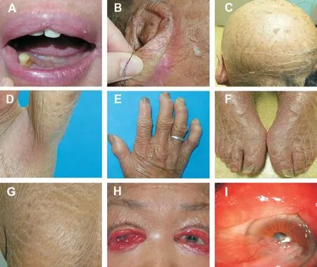
Clinical Features Proband 1 is an elderly patient with the chief complaint of binocular red eyes with tears since childhood and bad vision in the right eye for 2mo. Her bestcorrected visual acuity (BCVA) was light perception (LP)on the right and 0.6 on the left with the Snellen chart. The intraocular pressure was normal in both eyes. Proband 1 presented with severe upper and lower eyelid ectropion with lagophthalmos, absence of lacrimal punctum, deformity of the inner canthus, epithelialization of conjunctiva with severe hyperaemia and oedema, positive staining of the corneal surface with fluorescein and break-up time (BUT) <5s in both eyes. The fundus cannot be observed in the right eye and was essentially normal in the left eye. Bulbar conjunctiva hyperaemia and a 6×6-mm
white infiltration lesion were observed in the right eye. The lower half of the cornea and conjunctiva formed a symblepharon, and lenticular opacity was observed in the left eye (Figure 2).
The principles of treatment are to release the cicatrix and address the vertical deficiency of the anterior lamella by tailoring the graft to the individual patient
. The authors aim to enable the eyelids to close so that the ocular surface can be protected, and the appearance can be improved. These three probands had rare ED with classical ichthyosis-like skin changes, such as xeroderma, adiaphoresis, hyperkeratosis and desquamating. They lack healthy skin for transplantation, and there is a high possibility of contracture and poor healing of the grafting skin flap, which can easily lead to flap necrosis and recurrence of ectropion.
Detailed medical histories of the 3 probands were recorded.Peripheral venous blood was taken from 3 probands and their family members, and deoxyribonucleic acid (DNA) was extracted. Whole exome sequencing (WES) was conducted by the Beijing iGeneTech Institute (BII, China) using DNA from patients to identify potential pathogenic mutations. The authors filtered all the nonsynonymous single nucleotide polymorphisms (SNPs; synonymous, missense, nonsense,and splicing mutations) and inDels (short coding insertions or deletions) based on a minor allele frequency (MAF) ≤0.01, in 1000 Genomes, Exome Aggregation Consortium (ExAC) and Genome Aggregation database. Variants that are not at exonic or splicing gene regions were removed.
Then according to the database of all the genes linked to ED and ocular diseases which had been published previously, and deleterious result in functional prediction website as Sorting Intolerant From Tolerant (SIFT) and PolyPhen -2 program, the known variants (present in databases of normal people) and the non-deleterious ones were removed. In this way, the number of candidate genes was reduced to nine (
,
,
,
,
,
,
,
and
)in proband 1; sixteen (
,
,
,
,
,
,
,
,
,
,
,
,
,
,
,
) in proband 2; two (
and
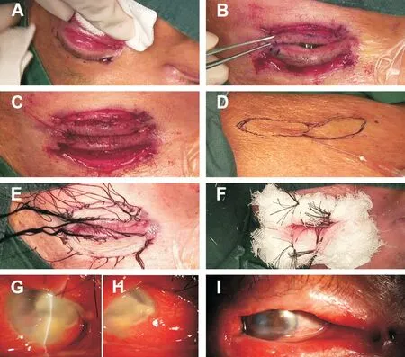
) in proband 3. These candidate pathogenic variants were amplified with polymerase chain reaction (PCR),and the PCR products were directly compared among the probands and their family members using Sanger sequencing.All the probands underwent surgery for free skin flap transplantation to correct ectropion. All surgeries were completed by one experienced surgeon. The surgical procedures were similar for both the upper and lower eyelids.After general anaesthesia, parallel incisions on the upper and lower lids were marked 3 mm away from the eyelid margin from the medial to the lateral cantus by methylene blue. After the eyelid was infiltrated with 1 to 2 mL of 2% lidocaine with 1:100 000 epinephrine, the incision was made meticulously along the marked line with a No.11 blade (Figure 1A).Westcott scissors were used to dissect the adhesive tissue until the eyelid margin was returned to its normal position without lagophthalmos (Figure 1B). Then, the anterior lamellar defects of the upper and lower eyelids were measured (Figure 1C). A full-thickness free graft was harvested from the preferred donor site, including the groin, abdomen and upper arm (Figure 1D). The graft was first thinned by trimming subcutaneous fat using Westcott scissors, followed by further trimming to match the exact shape of the recipient site. The overlying eyelid skin and grafting skin were sutured with interrupted 5-0 silk sutures carefully (Figure 1E). The skin graft was perforated to allow drainage and was then compressed by pressure pads for at least 7d (Figure 1F). Proband 2 only underwent bilateral upper eyelid surgery because the position of their lower eyelid ectropion did not meet the requirements of the operation, and proband 3 only received left eye surgery due to monophthalmia. According to the ichthyosis-like skin changes of these 3 patients, the surgeon chose relatively normal skin to transplant, which included the skin of the groin, abdomen and upper arm. Proband 3 was treated with bilateral nasal dacryocystorhinostomy under nasal endoscopy after ectropion surgery because of her chronic dacryocystitis in both eyes. All probands were followed up for at least one year to evaluate the results of the surgeries.
RESULTS
Clinical data of 3 probands diagnosed with ED at Eye Hospital of Wenzhou Medical University between December 2017 and January 2019 were collected.
两个好吃嘴边吃边聊边走,吃得嘴唇冒油光,脸上都是那油竹扦蹭出来的印子,空荡荡的胃被填补起来,身边的一切也都变得温暖起来,炸串儿全都下了肚还意犹未尽,恨不得和猫咪一样把牙齿上残留着的渣渣都舔干净。如今想来,那种香喷喷的滋味真的再难寻得。
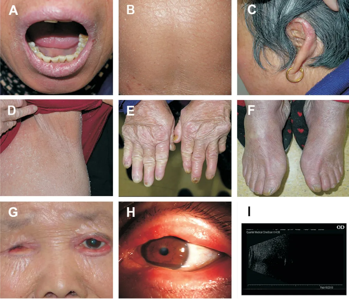
Proband 2 is a middle-aged patient with the chief complaint of binocular red eyes with tears and poor vision for more than 20y. Her BCVA was 0.16 on the right and 0.6 on the left with the Snellen chart. The intraocular pressure was normal in both eyes. Proband 2 presented with severe upper and lower eyelid ectropion with lagophthalmos, absence of lacrimal punctum, conjunctiva with hyperaemia and oedema,the corneal surface stained positive with fluorescein, and BUT<5s in both eyes. The fundus and lens were essentially normal in both eyes.

Regarding systemic manifestations, proband 2 had sweat gland manifestations similar to those of proband 1. Her teeth were essentially normal. There was no normal hair covering her whole body. In addition, proband 2 lacked eyebrows and eyelashes, though her scalp hair texture was normal. Her fingernails and toenails were abnormally shaped. She had skin pigmentation of the limbs, and other skin changes were similar to those of proband 1. Her face and hand skin were artificially smooth and lacked scales due to personal nursing. In addition,proband 2 also had finger joint contracture, dysplastic ears and frontal bossing (Figure 3).
Proband 3 is an elderly patient with the chief complaint of bad eye vision of the left eye for more than 10y. Her BCVA was non-light perception on the right and counting fingers (CF)on the left. The intraocular pressure was normal in the left eye. Proband 3 had congenital atrophy of the right eye, which was consistent with the B-scan ultrasonography results. She presented with severe upper and lower eyelid ectropion with lagophthalmos, absence of lacrimal punctum, conjunctiva with hyperaemia and oedema, a 1.5-mm-diameter central corneal nebula, the corneal surface stained positive with fluorescein and BUT<5s and lenticular opacity in the left eye. The fundus was essentially normal in the left eye.
Regarding systemic manifestations, proband 3 had sweat gland manifestations similar to those of proband 1. Her teeth were essentially normal. There was no normal hair covering her whole body. The scalp hair was essentially normal. Her fingernails and toenails were abnormally shaped, and some were blackened. Her skin was similar to proband 1, but her skin change was milder. In addition, proband 3 also had finger joint contracture, dysplastic ears and hearing impairment(Figure 4).
Table 1 shows the clinical data and systematic symptoms of the 3 probands. Table 2 summarizes the pre- and postoperative comparisons of ocular abnormalities of the 3 probands.
苏共长期执政,由于权力过分集中,缺乏监督制约,在苏共内部逐步形成了一个特殊的阶层。他们享有多方面特权:
Gene Sequencing There were no pathogenic gene mutations found in the three families. Proband 2 and her mother had the same heterozygous mutation in the
(c.871C>T,p.R291.W; NM_017410.2; Figure 5). Considering the clinical manifestations, their phenotypes are inconsistent; therefore,the mutation cannot explain the clinical manifestations and is not the pathogenic gene locus. For genetic patterns, in these three families, only the generation of probands had symptoms according to family pedigree. Proband 1 was born to non-consanguineous parents. Her older brother and halfsister presented similarly to her, but her elder brother died because of unknown high fever in childhood and her halfsister died in childhood with unknown reasons. Proband 2 was born to consanguineous parents. Proband 3 was born to nonconsanguineous parents. Her two older brothers presented similarly to her, but they died in childhood for unknown reasons. Except affected individuals above, there was no family history of skin, teeth, nails, sweat glands and other abnormalities in these three families’ members.
In the generation of probands of the first and third families,both males and females were patients. It is inferred that the genetic pattern might be autosomal recessive inheritance based on Mendel’s laws of inheritance. When both the normal father and mother carry a gene that causes the disease, the child is homozygous for the recessive gene and appears to have the disease. In the second family, the parents of proband 2 had a consanguineous marriage, which may increase the incidence of recessive genetic diseases. It has been reported that the risk of having congenital malformations doubles when a child is born in consanguineous marriage. Therefore, the genetic model of proband 2 might be an individual mutation or autosomal recessive in this family, and parental consanguineous marriage increases the probability of the disease.
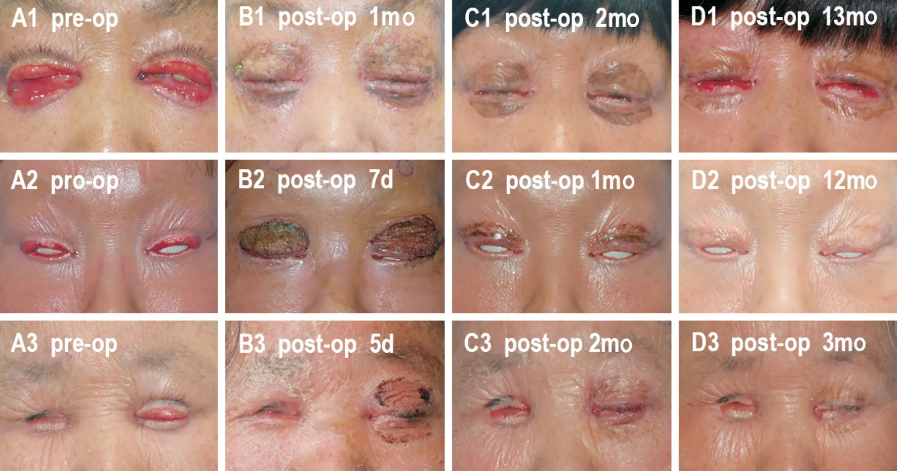
Surgery Outcomes During the postoperative 1-year followup period, the three probands all showed a good apposition in their eyelids (Figure 6). None of the grafts required removal or replacement. There were no graft complications of bleeding,haematoma formation, graft infection, hypertrophy or failure,except for one acceptable postoperative mild skin pigmentation in proband 1. The edges of the wound recovered well without hypertrophic scars. The incisions of the donor sites healed well.Symptoms of dry eye, eye pain and weeping improved. The BCVA of proband 1 was improved from preoperative LP to postoperative 0.2 in the right eye. The BCVA of proband 3 was improved from preoperative CF to postoperative 0.05 in the left eye. For proband 1, the 6×6-mm
white infiltration lesion on the cornea disappeared (Figure 1G-1I). For all probands,lagophthalmos, conjunctival hyperaemia and oedema, and fluorescence staining of the cornea were relieved.


DISCUSSION
In this study, three rare cases of ED with ophthalmopathy were reported. Their ocular clinical features include congenital eyelid defects, severe ectropion, hypophasis, absence of meibomian glands, deformity of the lacrimal duct (absence of lacrimal puncta), conjunctival epithelialization, keratopathy,cataracts, congenital monocular eyeball atrophy, and scarcity or absence of eyebrows and eyelashes. Genetic heterogeneity and diverse clinical manifestations lead to complex and diverse classifications. ED nosology always requires additions and modifications due to the identification of new genes and genetic alterations over time
. According to the Freire-Maia classification, three probands in this study should be classified in Group A, which has 163 subtypes. This study supplements the above subgroup classification and indicates that ectropion,lagophthalmos and absence of lacrimal punctum should be observed as key clinical traits of ED with ocular abnormality. ED with severe ectropion is very rare. Through a review of the literature,one case of secondary monocular ectropion due to dysplasia of the eyelid was reported. After ectropion blepharoplasty surgery,the patient’s right lower eyelid returned to the normal position, and no ectropion recurred during the 10-month follow-up period
.In this report, probands 1 and 3 were elderly women with cataracts. The researchers should not only consider agerelated cataracts but also note that the ectoderm is involved in lens development; therefore, it is likely that cataracts in these probands are more severe due to ED, which can be proven by the evaluation of more cases. In addition, there have been few studies that reported ED with symptoms of congenital eyeball atrophy showed in proband 3, which needs to be verified by more evidence.
For treatment, the common prominent ocular abnormality of the three probands was severe ectropion. When the lid skin is too tight vertically to allow the lid margin to be pulled up against the globe
, poor eyelid closure and the blinking could lead to corneal exposure, inadequate tear film distribution,and chronic ocular surface irritation
. Because of continued exposure, the palpebral conjunctiva frequently shows infiltrative and degenerative changes and may be considerably thickened
. Corneal damage arising from ectropion can be eased with moisturizers and artificial tears eye drop, while chronic inflammation can be eased medically with steroids and immunomodulators. As a result, surgery is needed to cure advanced ectropion by reconstructing the anterior lamellar defect of the eyelids.
其次,企业内部与银行建立信息交互机制,让金融机构与企业进行信息共享,提高自身信用难度。优化企业自身的资产信用度是解决餐饮企业融资的有效方式。
控制变量。包括企业规模、资产收益率、资产负债率、产权性质、上市时间,并进一步控制了年份和行业影响。控制变量的选取参照了Aihua-Wu等 (2016);郭晓丹等 (2011) 的研究。
There are reports citing the use of autografts from the arm, ipsi- or contralateral eyelid
, re-auricular or retro-auricular region, groin or penile foreskin in males
, human-engineered skin, mucous membrane grafts and maternal skin grafts (which require human leukocyte antigen typing and carry the risk of graft rejection) for reconstructing the eyelid skin
. According to the patients’ skin condition, the surgeon should select relatively normal skin to repair eyelid defects. The results within the followup period show that ectropion correction with full-thickness free skin flap transplantation is an effective procedure that restores the normal eyelid anatomical position and effectively relieves eye surface symptoms. Ectropion correction operations provide good ocular surface conditions for patients to undergo further ophthalmic surgery, such as cataract surgery and symblepharon separation. The authors will continue to observe the long-term recovery effect of these surgeries.
For genetic analysis, the authors did not detect any meaningful gene mutation and cannot establish the correlation of this mutation with the clinical manifestations of the patients.Although the genetic mechanism of the disease in the three families could not be clarified in this study, the similarity in key clinical traits may suggest an involvement of a specific underlying functional pathway. Identifying these various relationships may also suggest directions for future research.This study can be used as a reference for future research if they are verified in larger sample families.
Considering the rarity of ED, the study sample was small;therefore, it was difficult to obtain effective statistical results.Since there is no specialized grading scale for ectropion correction, the authors use related objective evaluation indicators for reference, such as the cornea, static asymmetry and dynamic and synkinesis (CADS) score
. The followup duration could be longer to allow us to indicate the median time to recurrence. The genetic sequencing did not yield positive results, and the patients lacked molecular diagnoses.The underlying genetic abnormalities, molecular pathways and the correlation of gene mutation with the clinical manifestations of the patients need to be elucidated in large samples of the family.
In summary, ectropion, lagophthalmos and the absence of lacrimal punctum might be three important ocular manifestations of ED, and ectropion needs to be relieved by surgery promptly. According to one-year postoperative observations, full-thickness free skin flap grafting is an effective surgery to correct ectropion in ED patients with ichthyosis-like skin changes. Future studies with longer followup periods are required to evaluate the long-term effectiveness of the functional and aesthetic outcomes in patients with ED.The suspected pathogenic genes of ED with ectropion should be further verified or confirmed by large samples of the family.
ACKNOWLEDGEMENTS
Authors’ contributions: Conception or design of the work:Zhang ZD, Chen X; Data collection: Duan BY, Zeng WX, Lin YY, Liu J; Data analysis and interpretation: Zhang ZD, Chen X, Duan BY, Zeng WX; Drafting the article: Zeng WX, Duan BY; Critical revision of the article: Zhang ZD, Chen X; Final approval of the version to be published: Zhang ZD, Chen X,Zeng WX, Duan BY, Lin YY, Liu J.
取40 μL经过稀释的乳液样品滴在载玻片上,小心盖上盖玻片,确保盖玻片和载玻片中间没有气泡。用奥林巴斯光学显微镜观察乳液的显微结构(目镜10倍,物镜40倍)。将制备好的样品放置在显微镜下观察,物镜放大10和20倍数,找到有代表性的物象后,用显微镜附带的相机拍照。
Foundation: Supported by Zhejiang Provincial Natural Science Foundation of China (No.LY19H120005).Conflicts of Interest: Chen X, None; Zeng WX, None; Duan BY, None; Lin YY, None; Liu J, None; Zhang ZD, None.
1 Wright JT, Fete M, Schneider H,
. Ectodermal dysplasias:classification and organization by phenotype, genotype and molecular pathway.
2019;179(3):442-447.
2 García-Martín P, Hernández-Martín A, Torrelo A. Ectodermal dysplasias: a clinical and molecular review.
2013;104(6):451-470.
3 Freire-Maia N. Ectodermal dysplasias revisited.
1977;26(2):121-131.
4 Pagnan NAB, Visinoni ÁF. Update on ectodermal dysplasias clinical classification.
2014;164A(10):2415-2423.
5 Priolo M, Laganà C. Ectodermal dysplasias: a new clinical-genetic classification.
2001;38(9):579-585.
6 Okubo A, Fujii K, Arimura A, Katsue H, Higashi Y, Shimomura Y,Kanekura T. Hypohidrotic ectodermal dysplasia with strabismus.
2018;45(7):e191-e192.
7 Wolosin JM, Budak MT, Akinci MAM. Ocular surface epithelial and stem cell development.
2004;48(8-9):981-991.
8 Kennedy DP, Chandler JW, McCulley JP. Ocular surface involvements in ectrodactyly-ectodermal dysplasia-cleft syndrome.
2015;38(3):228-231.
9 Koubek M, Strakošová K, Timkovič J, Grečmalová D, Orlíková A, Burčková H, Wiedermannová H, Mašek P. A rare form of ankyloblepharon filiforme adnatum associated with the Hay-Wells syndrome and a c.1709T>C mutation on the TP63 gene.
2018;39(2):251-254.
10 Ohdo S, Hirayama K, Terawaki T. Association of ectodermal dysplasia,ectrodactyly, and macular dystrophy: the EEM syndrome.
1983;20(1):52-57.
11 Djalilian AR, Kim JY, Saeed HN, Holland EJ, Chan CC. Histopathology and treatment of corneal disease in keratitis, ichthyosis, and deafness(KID) syndrome.
2010;24(4):738-740.
12 Callea M, Vinciguerra A, Willoughby CE, Deroma L, Clarich G.Infantile bilateral glaucoma in a child with ectodermal dysplasia.
2013;34(1-2):58-60.
13 Mukkamala K, Gentile RC, Willner J, Tsang S. Choroideremia in a woman with ectodermal dysplasia and complex translocations involving chromosomes X, 1, and 3.
2010;31(4):178-182.
14 Grogg JA, Port N, Graham T. Retinal tear presenting in a patient with ectrodactyly ectodermal dysplasia.
2014;91(4 Suppl 1):S55-S60.
15 Huffman RI, Huang JJ. Subretinal fibrosis and uveitis syndrome in a patient with ectodermal dysplasia.
2009;17(5):348-350.
16 Felipe AF, Abazari A, Hammersmith KM, Rapuano CJ, Nagra PK,Peiro BM. Corneal changes in ectrodactyly-ectodermal dysplasiacleft lip and palate syndrome: case series and literature review.
2012;32(5):475-480.
17 di Iorio E, Kaye SB, Ponzin D, Barbaro V, Ferrari S, Böhm E,Nardiello P, Castaldo G, McGrath JA, Willoughby CE. Limbal stem cell deficiency and ocular phenotype in ectrodactyly-ectodermal dysplasia-clefting syndrome caused by
mutations.
2012;119(1):74-83.
18 Zhang XY, Xu L, Li XF, Li CY, Zhang HT. Lower lid ectropion in hypohidrotic ectodermal dysplasia.
2015;2015:952834.
19 Karadağ R, Sevimli N, Karadağ AS, Wollina U. Successful correction of ichthyosis-related ectropion by autografts.
2020;33(6):e13851.
20 Hakim F, Phelps PO. Entropion and ectropion.
2020;66(10):101039.
21 Frueh BR, Schoengarth LD. Evaluation and treatment of the patient with ectropion.
1982;89(9):1049-1054.
22 Cannon PS, Madge SN, Kakizaki H, Selva D. Composite grafts in eyelid reconstruction: the complications and outcomes.
2011;95(9):1268-1271.
23 Mol I, Paridaens D. Efficacy of lateral eyelid-block excision with canthoplasty and full-thickness skin grafting in lower eyelid cicatricial ectropion.
2019;97(4):e657-e661.
24 Nayak S, Rath S, Kar BR. Mucous membrane graft for cicatricial ectropion in lamellar ichthyosis: an approach revisited.
2011;27(6):e155-e156.
25 Jen M, Nallasamy S. Ocular manifestations of genetic skin disorders.
2016;34(2):242-275.
26 Maqsood SE, Cascone N, Grixti A, Kannan R, Nduka C, Malhotra R. Functional and aesthetic outcomes of eyelid skin grafting in facial nerve palsy.
2018.
猜你喜欢
天天爱科学(2022年4期)2022-11-08
新作文·小学高年级版(2020年3期)2020-07-09
黄河黄土黄种人(2019年6期)2019-07-15
学苑创造·C版(2017年12期)2018-01-17
优雅(2017年11期)2017-11-11
儿童故事画报·发现号趣味百科(2016年7期)2017-02-08
优雅(2016年2期)2016-06-03
宠物世界·猫迷(2016年3期)2016-04-23
小学阅读指南·低年级版(2015年2期)2015-03-17
小学生·多元智能大王(2014年9期)2014-08-28
 International Journal of Ophthalmology2022年7期
- International Journal of Ophthalmology的其它文章
- Impact of OCT scan-patterns in identifying morphological features of lamellar macular holes and macular pseudoholes
- Virtual reality training improves accommodative facility and accommodative range
- Short-term effect of 0.01% atropine sulphate eye gel on myopia progression in children
- Reduced choroidal peripapillary capillaries in thyroidassociated ophthalmopathy with early stage of dysthyroid optic neuropathy
- Incidence of ocular manifestations in patients with graft versus host disease after allogeneic stem cell transplant in Riyadh, Saudi Arabia
- Temporal retinal thinning might be an early diagnostic indicator in male pediatric X-linked Alport syndrome