
锚蛋白重复序列22(ANKRD22)对人肝癌细胞的影响及其机制
2024-06-06 10:09蔡浚哲刘松柏费晓斌刘鹏朱昌毫王兴潘耀振
临床肝胆病杂志 2024年5期
蔡浚哲 刘松柏 费晓斌 刘鹏 朱昌毫 王兴 潘耀振

摘要: 目的 探討锚蛋白重复序列22(ANKRD22)对人肝癌细胞增殖、 侵袭和迁移的影响及其分子机制。方法 通过TCGA数据库分析正常肝组织及肝细胞癌组织中ANKRD22的表达水平及其与预后的关系。通过qRT-PCR和Western Blot检测人正常肝细胞 (L-02) 和人肝癌细胞系 (Huh7、 Hep G2、 MHCC-97H、 SK-HEP-1、 SMMC-7721) 中ANKRD22的表达情况。通过CCK-8、 EdU、 划痕实验及Transwell检测ANKRD22对肝癌细胞增殖、 侵袭和迁移能力的影响。通过Western Blot检测ANKRD22与细胞周期蛋白、 EMT相关蛋白之间的关系。通过KEGG、 ssGSEA分析进一步探究ANKRD22在肝癌细胞中的作用机制, 并进行实验验证。计量资料两组间比较采用成组t检验, 多组间比较采用单因素方差分析, 进一步两两比较采用LSD-t检验。结果 TCGA数据库中ANKRD22在肝细胞癌组织中较正常肝组织高表达 (t=5. 083, P<0. 05), 且ANKRD22高表达患者的总生存期及疾病相关生存期均显著低于ANKRD22低表达的患者 (P值均<0. 05)。肝癌细胞系中ANKRD22的表达量均高于正常肝细胞 (P值均<0. 05)。增殖实验结果提示, ANKRD22过表达组的EdU阳性率、 增殖速度均高于空载对照 (Vector) 组 (t值分别为19. 60、 6. 72, P值均<0. 001); si-ANKRD22#2组及si-ANKRD22#3组的EdU阳性率、 增殖速度较si-NC组均降低 (P值均<0. 001)。Cyclin E1、 Cyclin D1、 CDK7、 CDK4在过表达组中表达高于Vector组 (t值分别为3. 54、 4. 95、6. 34、 5. 19, P值均<0. 01); 在si-ANKRD22#2组及si-ANKRD22#3组中表达均低于si-NC组 (P值均<0. 001)。P27在过表达组中表达低于 Vector 组(t=6. 12, P<0. 001), 在 si-ANKRD22#2 组及 si-ANKRD22#3 组中表达均高于 si-NC 组(P 值均<0. 001)。侵袭、 迁移实验结果提示, ANKRD22过表达组的迁移速度、 穿膜数量 (迁移组和侵袭组) 均高于Vector组 (t值分别为5. 01、 25. 60、 3. 67, P值均<0. 05); si-ANKRD22#2组及si-ANKRD22#3组的迁移速度、 穿膜数量 (迁移组和侵袭组) 较si-NC组均降低 (P值均<0. 01)。N-cadherin、 Vimentin、 Snail在过表达组中表达高于Vector组 (t值分别为12. 13、 8. 85、 13. 97,P值均<0. 001), 在si-ANKRD22#2组及si-ANKRD22#3组中表达均低于si-NC组 (P值均<0. 001); E-cadherin在过表达组中表达低于Vector组 (t=4. 98, P<0. 01), 在si-ANKRD22#2组及si-ANKRD22#3组中表达均高于si-NC组 (P值均<0. 001)。KEGG富集分析及ssGSEA分析提示, ANKRD22在肝细胞癌中与PI3K/AKT/mTOR信号通路相关, 在过表达组中, p-AKT/AKT、p-PI3K/PI3K、 p-mTOR/mTOR 均较 Vector 组升高(t 值分别为 12. 21、 3. 43、 9. 75, P 值均<0. 01); 在 si-ANKRD22#2 组及si-ANKRD22#3组中表达均低于si-NC组 (P值均<0. 001)。结论 ANKRD22在肝癌细胞中高表达, 能促进肝癌细胞的增殖、侵袭和迁移能力, 并且能促进PI3K/AKT/mTOR信号通路的激活。
关键词: 癌, 肝细胞; 锚蛋白重复; 细胞增殖; 细胞运动
基金项目: 国家自然科学基金 (81960431, 81960535); 贵州省科技计划项目 (黔科合支撑 〔2021〕 一般444)
Effect of ankyrin-repeat domain-containing protein 22 on human hepatoma cells and its mechanism
CAI Junzhe1, LIU Songbai2, FEI Xiaobin1, LIU Peng3, ZHU Changhao4, WANG Xing1, 4, PAN Yaozhen1, 4.(1. College of Clinical Medicine, Guizhou Medical University, Guiyang 550000, China;2. Department of Hepatobiliary Surgery, The Affiliated Baiyun Hospital of Guizhou Medical University, Guiyang 550000, China; 3. Department of Hepatobiliary Surgery, The Affiliated Hospital of Guizhou Medical University, Guiyang 550000, China; 4. Department of Hepatobiliary Surgery, The Affiliated Cancer Hospital of Guizhou Medical University, Guiyang 550000, China)
Corresponding author: PAN Yaozhen, panyaozhen@gmc.edu.cn (ORCID: 0000-0002-9300-382X)
Abstract:
ObjectiveTo investigate the effect of ankyrin-repeat domain-containing protein 22 (ANKRD22) on the proliferation, invasion, and migration of human hepatoma cells and its molecular mechanism. Methods The TCGA database was used to analyze the expression level of ANKRD22 in normal liver tissue and hepatocellular carcinoma tissue and its association with prognosis. Western Blot and qRT-PCR were used to measure the expression of ANKRD22 in human normal liver cells (L-02) and human hepatoma cells (Huh7, HepG2, MHCC-97H, SK-HEP-1, and SMMC-7721); CCK-8 assay, EdU, wound healing assay, and Transwell assay were used to observe the effect of ANKRD22 on the proliferation, invasion, and migration of hepatoma cells; Western Blot was used to investigate the association of ANKRD22 with cyclins and EMT-related proteins; KEGG and ssGSEA analyses were performed to investigate the mechanism of action of ANKRD22 in hepatoma cells, and related experiments were conducted for validation. The independent-samples t-test was used for comparison of continuous data between two groups; a one-way analysis of variance was used for comparison between multiple groups, and the least significant difference t-test was used for further comparison between two groups. Results In the TCGA database, the expression level of ANKRD22 in hepatoma tissue was significantly higher than that in normal liver tissue (t=5.083, P<0.05), and the patients with a high expression level of ANKRD22 had longer overall survival and disease-related survival than those with a low expression level of ANKRD22 (P<0.05) . The expression level of ANKRD22 in various human hepatoma cell lines was higher than that in human normal liver cells (all P<0.05) . Cell proliferation assay showed that the ANKRD22 overexpression group had significantly higher EdU positive rate and proliferation rate than the Vector group (t=19.60 and 6.72, both P<0.001), and compared with the si-NC group, the si-ANKRD22#2 group and the si-ANKRD22#3 group had significantly lower EdU positive rate and proliferation rate (all P<0.001) . Compared with the Vector group, the overexpression group had significantly higher expression levels of Cyclin E1, Cyclin D1, CDK7, and CDK4 (t=3.54, 4.95, 6.34, and 5.19, all P<0.01), and the si-ANKRD22#2 group and the si-ANKRD22#3 group had significantly lower expression levels than the si-NC group (all P<0.001) . The overexpression group had a significantly lower expression level of P27 than the Vector group (t=6.12, P<0.001), and the si-ANKRD22#2 group and the si-ANKRD22#3 group had a significantly higher expression level than the si-NC group (both P<0.001) . Invasion and migration experiments showed that compared with the Vector group, the ANKRD22 overexpression group had significantly higher migration rate and number of crossings through the membrane (migration group and invasion group) (t=5.01,25.60, and 3.67, all P<0.05), and compared with the si-NC group, thesi-ANKRD22#2 group and the si-ANKRD22#3 group had significantly lower migration rate and number of crossings through the membrane (migration group and invasion group)(all P<0.01) . The overexpression group had significantly higher expression levels of N-cadherin, Vimentin, and Snail than the Vector group (t=12.13, 8.85, and 13.97, all P<0.001), and the si-ANKRD22#2 group and the si-ANKRD22#3 group had significantly lower expression levels than the si-NC group (all P<0.001); the overexpression group had a significantly lower expression level of E-cadherin than the Vector group (t=4.98, P<0.01), and the si-ANKRD22#2 group and the si-ANKRD22#3 group had a significantly higher expression level than the si-NC group (both P<0.001) . The KEGG enrichment analysis and the ssGSEA analysis showed that ANKRD22 was associated with the PI3K/AKT/mTOR signaling pathway in hepatocellular carcinoma, and the overexpression group had significantly higher expression levels of p-AKT/AKT, p-PI3K/PI3K, and p-mTOR/mTOR than the Vector group (t=12.21, 3.43, and 9.75, all P<0.01), and the si-ANKRD22#2 group and the si-ANKRD22#3 group had significantly lower expression levels than the si-NC group (all P<0.001) . Conclusion ANKRD22 is highly expressed in hepatoma cells and can promote the proliferation, invasion, and migration of hepatoma cells and the activation of the PI3K/AKT/mTOR signaling pathway.
Key words: Carcinoma, Hepatocellular; Ankyrin Repeat; Cell Proliferation; Cell Movement
Research funding: National Natural Science Foundation of China (81960431, 81960535); Science and Technology Fund of Guizhou Province (QKHZC [2021] YB444)
肝细胞癌 (HCC) 是世界上第六常见的恶性肿瘤, 并且是全球癌症死亡第四大相关因素[1] , 并逐渐呈现上升趋势[2] 。HCC在早期可无症状, 确诊时常已发展至中晚期[3] , 而晚期HCC患者可选择的治疗方案十分有限[4] 。尤其在我国, 相关药物仍未能满足临床上的诸多需求[5] 。因此, 继续寻找HCC相关的生物标志物具有重要价值。
锚蛋白重复序列22 (ankyrin-repeat domain-containing protein 22, ANKRD22)属于锚蛋白(ANK)重复序列家族[6] , 是一种人N-肉豆蔻酰化蛋白[7] , 含有4个重复的锚蛋白基序, 共有191个氨基酸, 并且在氨基酸87和109之间具有假定的单通道跨膜区域[8] 。ANKRD22在多种恶性肿瘤中高表达, 但其在HCC中的作用尚不明确。本研究通过对ANKRD22进行过表达或者敲低, 来分析其对人HCC细胞增殖、 侵袭和迁移的影响及其分子机制。
1 材料与方法
1. 1 实验材料 人正常肝细胞株L-02及肝癌细胞株Huh7、 Hep G2、 MHCC-97H、 SK-HEP-1和SMMC-7721均购于中国科学院上海分院细胞库; DMEM培养基、 0. 25%胰蛋白酶、 10%胎牛血清等均购于美国Gibco公司; 过表达慢病毒购于中国上海吉凯基因公司; siRNA购于中国广州锐博生物科技有限公司; Lipo3000 购于美国invitrogen公司; ANKRD22及相关抗体均购于中国武汉三鹰公司。
1. 2 生物信息学分析 在TCGA数据库 (https: //portal.gdc. cancer. gov/) 中下载ANKRD22在正常肝组织及肝细胞癌组织中的相关数据, 运用R4. 2. 1软件, 以 “limma” 和“edgR” R包进行差异表达分析, 以 “survival” 和 “survminer” R包进行预后分析。然后将TCGA数据库中筛选出的在HCC中与ANKRD22相关的基因进行KEGG富集分析寻找较多富集的通路, 并通过ssGSEA进行进一步分析。
1. 3 细胞培养 所有细胞株均使用含10%胎牛血清的DMEM培养基来进行培养, 细胞培养的环境为恒温37 ℃、含5% CO2的无菌培养箱, 待细胞培养至对数期生长时提取以进行后续进一步实验。
1. 4 细胞siRNA转染 将对数期生长的细胞Huh7取2×10⁵个接种于六孔板, 在细胞密度增至30%~40%时以Lipo3000为介质开始siRNA转染。先以无血清培养基将Lipo3000及siRNA分别孵育5 min, 然后进行混合孵育15 min, 孵育结束后缓慢滴入六孔板中并摇匀, 放入培养箱培养6 h后更换培养基, 24~48 h后可提取RNA并通过qRT-PCR验证siRNA转染效率, 48~72 h后可提取蛋白并通过Western Blot验证siRNA转染效率。
1. 5 细胞慢病毒感染 将对数期生长的细胞MHCC-97H取1×10⁵个接种于培养瓶内, 约24 h后开始病毒感染, 依照病毒的MOI值将病毒吸入培养瓶内并摇匀, 放入培养箱24 h后更换培养基, 约48 h后可提取RNA并通过qRT-PCR验证病毒转染效率, 约72 h后可提取蛋白并通过Western Blot验证病毒转染效率。
1. 6 qRT-PCR实验 收集待处理的细胞, 使用Trizol试剂以提取细胞的总RNA, 使用PrimeScriptTM RT Reagent试剂盒进行逆转录以得到cDNA, 使用SYBR?Premix Ex TaqTM试剂盒进行qRT-PCR分析, 每组分别设置5个复孔,反应条件为95 ℃、 30 s, 95 ℃、 5 s, 60 ℃、 30 s, 扩增40个循环, 得到的结果使用Bio-Rad CFX Manager进行分析, 以GAPDH基因作为内参, 使用公式2?ΔΔCt计算得到目的基因相对于GAPDH的表达量。
1. 7 Western Blot实验 收集待处理的细胞, 加入裂解液以提取细胞的总蛋白, 使用SDS-PAGE凝胶进行电泳以分离蛋白, 在300 mA下转膜至PVDF膜上, 用5%脱脂牛奶室温封闭2 h, 放入一抗4 ℃孵育过夜, 第二天放入二抗中室温孵育2 h, 滴加显影液后在化学发光仪中显影。
1. 8 划痕实验 将对数期生长的细胞取5×10⁵个接种于六孔板内, 待细胞覆盖大于90%孔径时, 以200 ?L规格枪头垂直划线, 并用PBS清洗后置于显微镜下拍照,放入培养箱中培养48 h后再次拍照。
1. 9 CCK-8实验 将对数期生长的细胞取3×10³个接种于96孔板内, 待细胞贴壁后加入CCK-8试剂, 计为0 h,并用酶标仪检测于450 nm处各孔吸光度值, 后分别于培养24 h、 48 h、 72 h、 96 h后重复检测。
1. 10 EdU增殖实验 将对数期生长的细胞取3×10⁴个细胞接种于24孔板内, 待细胞貼壁后加入EdU工作液于培养箱内孵育2 h, 然后弃去培养基、 固定细胞, 用TritonX-100透膜后加入荧光染料进行染色, 待染色结束后用荧光显微镜进行观察计数并计算阳性率。
1. 11 Transwell试验 将对数期生长的细胞悬浮于无血清培养基中, 将含3×10⁴个细胞的细胞悬液滴加于含或不含基质胶的上室内, 并将含20%胎牛血清的培养基滴加于下室内, 置于培养箱内培养24~48 h后吸去培养基, 用PBS清洗后加入4%多聚甲醛固定, 固定完成后用PBS清洗, 再加入0. 3%结晶紫固定, 染色结束后用PBS清洗, 待烘干后置于显微镜下观察细胞数量。
1. 12 统计学分析 使用SPSS 25. 0统计软件进行分析, 计量资料两组间比较采用成组t检验, 多组间比较采用单因素方差分析, 进一步两两比较使用LSD-t检验, P<0. 05为差异具有统计学意义。
2 结果
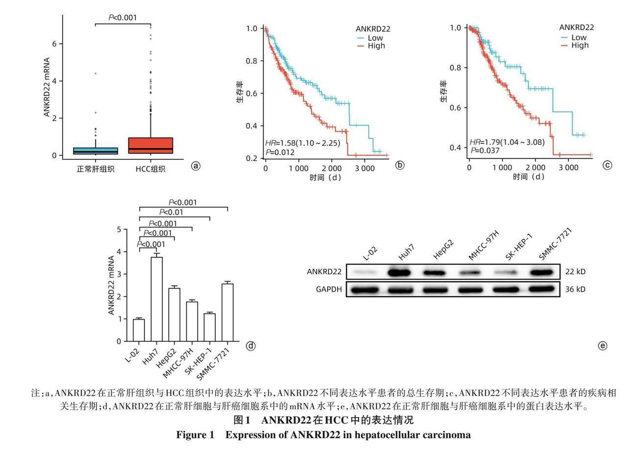
2. 1 ANKRD22在HCC中的表达 通过对TCGA数据库进行分析, 结果提示ANKRD22在HCC组织中较正常肝组织高表达 (t=5. 083, P<0. 001)(图1a), 且ANKRD22高表达患者的总生存期及疾病相关生存期均显著低于低表达的患者 (P值均<0. 05)(图1b、 c)。进一步以qRT-PCR及Western Blot对肝癌细胞系中ANKRD22表达情况进行检测, 结果提示肝癌细胞系中ANKRD22的表达量均高于正常肝细胞 (P值均<0. 05)(图1d、 e); 其中Huh7的表达量最高, MHCC-97H的表达量次之, 故以此2种细胞进行后续实验。
2. 2 ANKRD22转染后的表达情况 使用过表达慢病毒转染 MHCC-97H, 使用 siRNA 转染 Huh7, 并以 qRT-PCR 及 Western Blot 进行验证。结果提示, 过表达组MHCC-97H的ANKRD22 mRNA (t=85. 21, P<0. 001) 及蛋白(t=35. 27, P<0. 001) 表达量均明显高于空载对照组 (Vector组)(图2a、 b); 3个敲低组Huh7的mRNA及蛋白表达量均较阴性对照组 (si-NC组) 降低 (P值均<0. 01), 其中si-ANKRD22#2组及si-ANKRD22#3组的敲低效果较为显著(图2c、 d), 故继续以si-ANKRD22#2及si-ANKRD22#3完成后续实验。以上结果提示过表达组和敲低组均构建成功。
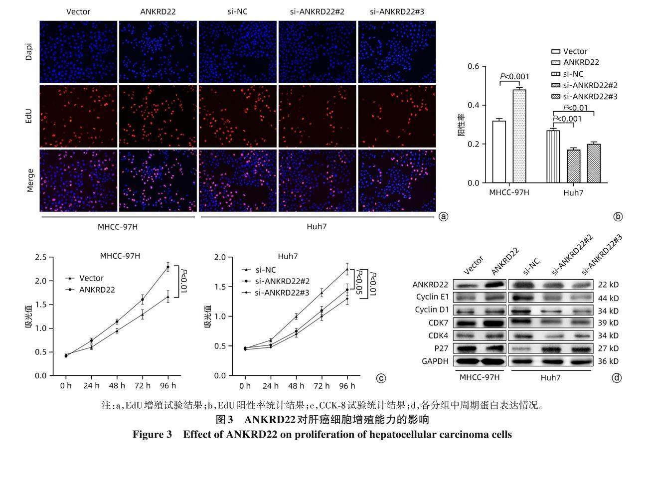
2. 3 ANKRD22对肝癌细胞增殖能力的影响 EdU增殖实验结果提示, 过表达组的阳性率高于 Vector 组(t=19. 60, P<0. 001); si-ANKRD22#2组及si-ANKRD22#3组的阳性率较si-NC组均降低 (P值均<0. 001)(图3a、 b)。CCK-8实验结果提示, 过表达组的增殖速度高于Vector组 (t=6. 72, P<0. 01); si-ANKRD22#2组及si-ANKRD22#3组的增殖速度均较si-NC组降低 (P值均<0. 05)(图3c)。Western Blot结果提示, Cyclin E1、 Cyclin D1、 CDK7、 CDK4在过表达组中表达高于Vector组 (t值分别为3. 54、 4. 95、 6. 34、5. 19, P值均<0. 01); 在si-ANKRD22#2组及si-ANKRD22#3组中表达均低于si-NC组 (P值均<0. 001)。P27在过表达组中表达低于Vector组 (t=6. 12, P<0. 001), 在si-ANKRD22#2组及si-ANKRD22#3组中表达均高于si-NC组 (P值均<0. 001)(图3d)。
2. 4 ANKRD22对肝癌细胞侵袭、 迁移能力的影响 划痕实验结果提示, 过表达组MHCC-97H的迁移速度高于Vector组 (t=5. 01, P<0. 01); si-ANKRD22#2组及si-ANKRD22#3组的迁移速度均较si-NC组降低 (P值均<0. 01)(图4a、b) 。Transwell实验结果提示, 过表达组的穿膜数量高于Vector组 (迁移组: t=25. 60, P<0. 001; 侵袭组: t=3. 67, P<0. 05); si-ANKRD22#2组及si-ANKRD22#3组的穿膜数量均较si-NC组降低 (P值均<0. 01)(图4c~e)。进一步通过Western Blot分别对Vector组、 过表达组、 si-NC组、 si-ANKRD22#2组、 si-ANKRD22#3组与EMT相关蛋白之间的关系进行验证, 结果提示N-cadherin、 Vimentin、 Snail在过表达组中表达高于Vector组 (t值分别为12. 13、 8. 85、13. 97, P值均<0. 001), 在si-ANKRD22#2组及si-ANKRD22#3组中表达均低于si-NC组 (P值均<0. 001); E-cadherin在过表达组中表达低于Vector组 (t=4. 98, P<0. 01), 在si-ANKRD22#2组及si-ANKRD22#3组中表达均高于si-NC组 (P值均<0. 001)(图4f)。
2. 5 ANKRD22在肝癌细胞中的作用机制 KEGG富集分析结果提示, ANKRD22在HCC组织中富集于PI3K/AKT/mTOR、 JAK/STAT、 NF-κB等多条信号通路, 其中在PI3K/AKT/mTOR信号通路上的富集最为显著 (图5a)。ssGSEA富集分析结果提示, ANKRD22在HCC组织中正向显著富集于PI3K/AKT/mTOR信号通路 (图5b)。通过Western Blot对ANKRD22在肝癌细胞中对AKT、 PI3K、mTOR的磷酸化水平进行验证, 结果提示在过表达组中,p-AKT/AKT、 p-PI3K/PI3K、 p-mTOR/mTOR均较Vector组升高 (t值分别为12. 21、 3. 43、 9. 75, P值均<0. 01); 在si-ANKRD22#2 組及 si-ANKRD22#3 组中, p-AKT/AKT、 p-PI3K/PI3K、 p-mTOR/mTOR 均较 si-NC组降低(P值均<0. 001)(图5c)。
3 讨论
2021年统计数据[9] 显示, 我国肝癌患者数量占全球近50%, 肝癌作为在我国恶性肿瘤中发病率第4位、 致死率第2位的疾病[10] , 仍需继续高度关注。
ANK是自然界中最常见的蛋白质基序之一, 广泛参与多种细胞过程[11]。ANKRD22作为ANK家族中的一员, 可能与人体内的多种生物学功能有关。当前相关研究表明ANKRD22与多种恶性肿瘤相关, 其中Liu等[12] 发现ANKRD22可以通过上调E2F1介导的MELK表达促进胶质瘤增殖、 迁移、 侵袭和上皮-间充质转化; Wu等[13] 发现ANKRD22可以通过调节NuSAP1表达激活Wnt/β-连环蛋白通路来促进乳腺癌; Yin等[14] 发现ANKRD22可以通过上调E2F1的转录来促进非小细胞肺癌的进展; Wu等[15] 发现ANKRD22可以通过调节Wnt/β-连环蛋白信号通路从而影响甲状腺癌细胞的生长和迁移。因此笔者假设ANKRD22在肝癌细胞中也能起到促癌作用, 并且通过相关验证发现ANKRD22的确对肝癌细胞的增殖、侵袭和迁移起促进作用。但是, Pan等[16] 发现ANKRD22通过与E-syt1合作从而促进结直肠癌的代谢重编程;Chen等[17]发现ANKRD22可以通过逆转PMN-MDSC的免疫抑制作用从而成为卵巢癌治疗的新靶点; Qiu等[18]发现ANKRD22可能在前列腺癌的进程中起负向作用。说明ANKRD22在人体中有着复杂的生物学功能, 在肝癌细胞中也有可能有着更多的生物学功能, 本研究发现ANKRD22与PI3K/AKT/mTOR信号通路相关, 但具体作用方式尚不明确。
本研究通过TCGA数据库发现ANKRD22在人HCC组织中高表达且与患者的生存时间呈负相关。随后通过细胞实验发现, 在肝癌细胞中, ANKRD22较正常肝细胞中高表达且ANKRD22的表达水平与肝癌细胞的增殖、 侵袭和迁移能力呈正相关。为进一步探究ANKRD22在肝癌細胞中的作用机制, 通过富集分析发现其与PI3K/AKT/mTOR信号通路相关, 并通过实验证明ANKRD22的表达量与AKT、 PI3K、 mTOR的磷酸化水平呈正相关, 笔者推测ANKRD22通过影响PI3K/AKT/mTOR信号通路从而促进肝癌的进展。此外, PI3K/AKT/mTOR信号通路在肝癌中与代谢重编程[19] 、 上皮-间充质转化[20] 、 脂质合成[21]等多种生物学功能相关, ANKRD22具体如何作用于PI3K/AKT/mTOR信号通路影响肝癌细胞的增殖、 侵袭和迁移,ANKRD22是否通过其他机制调控肝癌细胞生长等方面尚不明确, 仍需继续进行后续研究进一步探索。
利益冲突声明: 本文不存在任何利益冲突。
作者贡献声明: 蔡浚哲、 刘松柏、 费晓斌负责实验操作;刘鹏、 朱昌毫负责资料收集与数据分析; 王兴负责课题设计; 潘耀振指导撰写论文并最后定稿。
参考文献:
[1] LLOVET JM, KELLEY RK, VILLANUEVA A, et al. Hepatocellular carci?noma[J]. Nat Rev Dis Primers, 2021, 7(1): 6. DOI: 10.1038/s41572-020-00240-3.
[2] FOGLIA B, TURATO C, CANNITO S. Hepatocellular carcinoma: Lat?est research in pathogenesis, detection and treatment[J]. Int J Mol Sci, 2023, 24(15): 12224. DOI: 10.3390/ijms241512224.
[3] DING CM, HOU JF, TAO GW, et al. Early diagnosis and screening of hepatocellular carcinoma[J/OL]. Chin J Hepatic Surg Electron Ed, 2023, 12(1): 22-28. DOI: 10.3877/cma.j.issn.2095-3232.2023.01.005.
丁成明, 侯嘉丰, 陶光伟, 等. 肝细胞癌早期诊断和筛查[J/OL]. 中华肝脏外科手术学电子杂志, 2023, 12(1): 22-28. DOI: 10.3877/cma.j.issn.2095-3232.2023.01.005.
[4] KIM E, VIATOUR P. Hepatocellular carcinoma: Old friends and new tricks[J]. Exp Mol Med, 2020, 52(12): 1898-1907. DOI: 10.1038/s12276-020-00527-1.
[5] LIU XF, ZHANG J, YAO L, et al. Advances in targeted therapy com?bined with immunotherapy for advanced hepatocellular carcinoma[J]. J Clin Hepatol, 2022, 38(5): 992-997. DOI: 10.3969/j.issn.1001-5256.2022.05.004.
刘秀峰, 张珏, 姚琳, 等. 中晚期肝细胞癌靶向联合免疫治疗进展[J]. 临床肝胆病杂志, 2022, 38(5): 992-997. DOI: 10.3969/j.issn.1001-5256.2022.05.004.
[6] YANG SS. Evaluation of Clinical Significance of ANKRD22 in Colorec?tal Cancer[D]. Hangzhou: Zhejiang University, 2019. DOI: 10.27461/d.cnki.gzjdx.2019.001668.
杨赛赛. ANKRD22在结直肠癌细胞中表达意义的研究[D]. 杭州: 浙江大学, 2019.
[7] UTSUMI T, HOSOKAWA T, SHICHITA M, et al. ANKRD22 is an N-myristoylated hairpin-like monotopic membrane protein specifically local?ized to lipid droplets[J]. Sci Rep, 2021, 11(1): 19233. DOI: 10.1038/s41598-021-98486-8.
[8] WANG R, WU YH, ZHU Y, et al. ANKRD22 is a novel therapeutic tar?get for gastric mucosal injury[J]. Biomed Pharmacother, 2022, 147: 112649. DOI: 10.1016/j.biopha.2022.112649.
[9] ZHOU HN, LI YM. Conversion therapy for hepatocellular carcinoma[J/OL]. Chin J Hepatic Surg Electron Ed, 2022, 11(6): 542-547. DOI: 10.3877/cma.j.issn.2095-3232.2022.06.002.
周辉年, 李玉民. 肝癌转化治疗[J/OL]. 中华肝脏外科手术学电子杂志, 2022, 11(6): 542-547. DOI: 10.3877/cma.j.issn.2095-3232.2022.06.002.
[10] LU SL, YAO JN, YUAN GD, et al. Current status and prospect of clinical and basic research on conversion therapy for hepatocellular carcinoma[J]. Chin J Exp Surg, 2022, 39(10): 1837-1843. DOI: 10.3760/cma.j.cn421213-20220210-01043.
陆世鎏, 姚建妮, 袁观斗, 等. 肝癌转化治疗临床与基础研究现状与展望[J]. 中华实验外科杂志, 2022, 39(10): 1837-1843. DOI: 10.3760/cma.j.cn421213-20220210-01043.
[11] PARRA RG, ESPADA R, VERSTRAETE N, et al. Structural and en?ergetic characterization of the ankyrin repeat protein family[J]. PLoS Comput Biol, 2015, 11(12): e1004659. DOI: 10.1371/journal.pcbi.1004659.
[12] LIU X, ZHAO JL, WU Q, et al. ANKRD22 promotes glioma prolifera?tion, migration, invasion, and epithelial-mesenchymal transition by upregulating E2F1-mediated MELK expression[J]. J Neuropathol Exp Neurol, 2023, 82(7): 631-640. DOI: 10.1093/jnen/nlad034.
[13] WU YG, LIU HX, GONG YF, et al. ANKRD22 enhances breast can?cer cell malignancy by activating the Wnt/β -catenin pathway via modulating NuSAP1 expression[J]. Bosn J Basic Med Sci, 2021, 21(3): 294-304. DOI: 10.17305/bjbms.2020.4701.
[14] YIN J, FU WF, DAI L, et al. ANKRD22 promotes progression of non-small cell lung cancer through transcriptional up-regulation of E2F1[J]. Sci Rep, 2017, 7(1): 4430. DOI: 10.1038/s41598-017-04818-y.
[15] WU YG, CHEN WX, ZHANG B, et al. ANKRD22 knockdown sup?presses papillary thyroid cell carcinoma growth and migration and modulates the Wnt/β-catenin signaling pathway[J]. Tissue Cell, 2023, 84: 102193. DOI: 10.1016/j.tice.2023.102193.
[16] PAN TH, LIU JW, XU S, et al. ANKRD22, a novel tumor microenvironment-induced mitochondrial protein promotes metabolic reprogramming of colorectal cancer cells[J]. Theranostics, 2020, 10(2): 516-536. DOI: 10.7150/thno.37472.
[17] CHEN HH, YANG KQ, PANG LX, et al. ANKRD22 is a potential novel target for reversing the immunosuppressive effects of PMN-MDSCs in ovarian cancer[J]. J Immunother Cancer, 2023, 11(2): e005527. DOI: 10.1136/jitc-2022-005527.
[18] QIU YQ, YANG SS, PAN TH, et al. ANKRD22 is involved in the pro?gression of prostate cancer[J]. Oncol Lett, 2019, 18(4): 4106-4113. DOI: 10.3892/ol.2019.10738.
[19] TIAN LY, SMIT DJ, J?CKER M. The role of PI3K/AKT/mTOR signal?ing in hepatocellular carcinoma metabolism[J]. Int J Mol Sci, 2023, 24(3): 2652. DOI: 10.3390/ijms24032652.
[20] LI YH, YIN YL, HE Y, et al. SOS1 regulates HCC cell epithelial-mes?enchymal transition via the PI3K/AKT/mTOR pathway[J]. Biochem Biophys Res Commun, 2022, 637: 161-169. DOI: 10.1016/j.bbrc.2022.11.015.
[21] CHEN JX, CHEN JD, HUANG JX, et al. HIF-2α upregulation medi?ated by hypoxia promotes NAFLD-HCC progression by activating lipid synthesis via the PI3K-AKT-mTOR pathway[J]. Aging (Albany NY), 2019, 11(23): 10839-10860. DOI: 10.18632/aging.102488.
收稿日期:2023-10-13; 录用日期:2023-11-17
本文编辑:王莹
猜你喜欢
传染病信息(2022年6期)2023-01-12
昆明医科大学学报(2022年2期)2022-03-29
昆明医科大学学报(2022年1期)2022-02-28
医学信息(2016年36期)2017-02-23
右江医学(2016年4期)2017-01-05
医学信息(2016年29期)2016-11-28
课程教育研究·学法教法研究(2016年6期)2016-04-26
癌变·畸变·突变(2016年3期)2016-02-27
哈尔滨医药(2015年4期)2015-12-01
现代养生·下半月(2015年8期)2015-11-16
