
Multimodal imaging in Purtscher-like retinopathy associated with sarcoidosis: a case report
2022-05-15 05:40HlaKaoualInsHachichaIlhemMiliBoussenImneZhiouaBrahamMejdiBoukariRajaZhioua
鼻咽癌是一种较常见的恶性肿瘤,发生在鼻咽部位,有研究表明,大多数为低分化鳞癌,具有较高的恶性程度,发生广泛转移、局部浸润的可能性也比较高。肿瘤细胞可能对颅底造成破坏,进而对脑组织形成侵犯。另外,对于鼻咽癌来说,其发生淋巴结转移的机率也相对较高。由于鼻咽癌发病位置解剖结构特殊,周围重要的神经组织、血管等比较丰富,因此手术治疗有较大的局限[1-2]。鼻咽癌对放射治疗敏感性较高,所以一般采用放射治疗的方法,常规放射治疗效果一般,采用调强放射治疗,对靶区三维形状进行获取,减少对正常组织的照射剂量,能保护正常组织,减少副反应,并且提高治疗效果[3-4]。
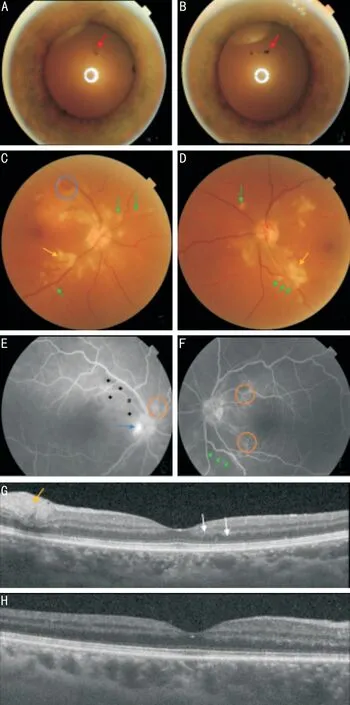
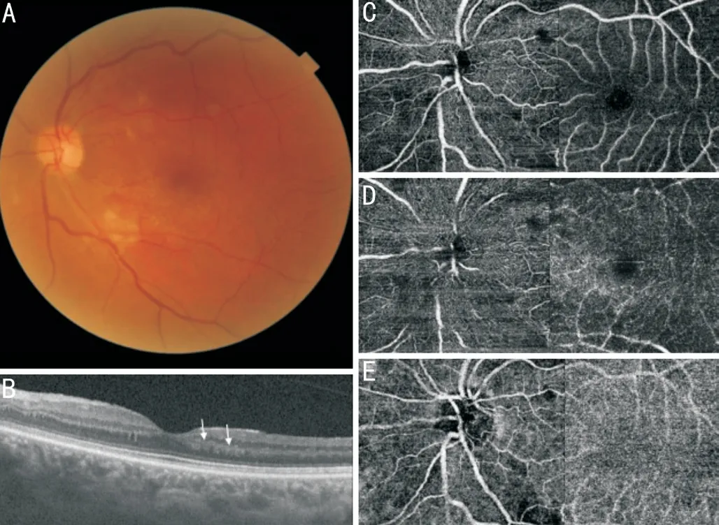
We report a case of Purtscher-like retinopathy (PLR)in the presence of acute pancreatitis secondary to sarcoidosis. To our knowledge, this is the first case report of a PLR in a patient with sarcoidosis. A 54-year-old female, hospitalized at the Internal Medicine Department for stage E pancreatitis, hypercalcemia, and mediastinal lymphadenopathies, consulted with a visual impairment in both eyes evolving for a month, especially in her left eye (LE). The best-corrected visual acuity (BCVA), was 20/32 in the right eye(RE) and 20/50 in the LE. The slit-lamp examination revealed iris pigments on the anterior lens capsule, corresponding to disrupted posterior synechiae in both eyes (Figure 1A,1B). The vitreous was clear, whereas fundus examination(Figure 1C, 1D) showed a stage 1 papillary edema and few superficial intra-retinal hemorrhages in the RE associated with multiple peri-papillary cotton-wool spots (CWS) in both eyes. Moreover, we noted focal whitening surrounding the retinal arterioles with a clear zone on either side of the vessels(Purtscher fleken). These findings were compatible with the diagnosis of PLR. Fluorescein angiography (FA) showed papillary early hyper fluorescence (papillary edema), peripapillary non-perfusion areas associated with late capillary staining. We also noted peri-papillary and peri-vascular hypofluorescent spots, corresponding respectively to CWS and Purtscher fleken (Figure 1E, 1F). Macular Swept-source optical coherence tomography (SS-OCT) detected a focal thickening of the retinal fiber layer corresponding to CWS in both eyes.Besides, hyperreflectivity of the inner nuclear layer consisting of a paracentral acute middle maculopathy (PAMM), was revealed in the LE (Figure 1G, 1H). However, no disruption of the ellipsoid zone or macular thickening was noted. Sweptsource optical coherence tomography angiography (SSOCTA) showed superficial and deep capillary non-perfusion(CNP) areas (Figure 2A-2D). Flow void areas were noted in the choriocapillaris, in addition to a shadowing effect due to the overlying CWS (Figure 2E, 2F). Regarding the association of ocular inflammation sequelae (disrupted posterior synechiae) with hypercalcemia and deep lymphadenopathies,the diagnosis of sarcoidosis was suspected. However, the dosage of angiotensin-converting enzyme and salivary gland biopsy was normal. Infectious causes, including tuberculosis,systemic diseases, and malignancies were ruled out. Thus,a mediastinoscope-guided biopsy of the lymphadenopathies was performed. Histopathological specimen revealed, giant cells with non-caseating granulomas, confirming the diagnosis of sarcoidosis. The patient received an immunosuppressive dose of systemic steroids, initiated by three intravenous boluses of methylprednisolone (1 g/d) followed by oral prednisolone(1 mg/kg·d). A month later, the patient’s vision improved, with a BCVA of 20/25 in both eyes. The fundus examination revealed a decrease in the number of CWS and the disappearance of retinal hemorrhages (Figure 3A). The PAMM persisted in the LE (Figure 3B). However, CNP and flow void areas decreased on OCTA (Figure 3C-3E).
此外,加之金融方式的介入以及B2C模式形成的在线直供,最终形成了一个完整的全产业链闭环。一位肥料企业老总“惊爆”:“传统肥料企业已经意识到迫近的形势,全产业链服务模式让我们没有活路了。”
DISCUSSION

Acute pancreatitis is the first cause of PLR, it represents 19.1% of cases
. The physiopathology of this retinopathy, in the cases of acute pancreatitis, was explained by complement activation that results in leukocyte aggregates or leukoemboli,which are released in the bloodstream and occlude the retinal vessels
. In sarcoidosis, pancreatic involvement is uncommon,moreover acute pancreatitis due to hypercalcemia is rare
. The association between systemic diseases and PLR was reported in some cases, however, to our knowledge, the association between sarcoidosis and PLR was never described. Sarcoidosis is well known to induce occlusive retinal vasculitis
, thus we hypothesized that microangiopathy that led to the PLR,in our case, could be explained by two vascular occlusive entities, sarcoidosis, and acute pancreatitis. In the context of a probable etiology, the diagnosis of PLR is based on a sudden visual impairment (bilateral in 60% of cases) and fundoscopic findings restricted to the posterior pole: CWS,retinal hemorrhages, and Purtscher flecken. Papillary edema has been observed in some cases
. FA shows different degrees of CNP areas and fluorescein staining from retinal vessels,according to the severity of the retinopathy. Hypo-fluorescent spots corresponding to the CWS and Purtscher flecken are also observed
. In the majority of cases of PLR, optical coherence tomography shows macular edema
. However, in this case, the macular thickness was normal, and a PAMM was revealed. This condition in the context of PLR, might be explained by the ischemia induced by the retinal arteriolar emboli
. SS-OCTA is a recent multimodal imaging technique that showed, in this case, not only multiple areas of CNP in the superficial and deep capillary plexuses but also flow void in the choriocapillaris, described by Li
as a honeycomblike pattern. This finding suggests the involvement of the choroid in the physiopathology of PLR. The evolution of PRL is favorable in some cases, however, no prognosis criteria have been fixed
. In this case, the patient regained a correct visual acuity in both eyes, despite the persistence of PAMM in her LE. This evolution might be explained by the decrease in CNP areas in the retinal capillary plexuses and the choriocapillaris as shown on SS-OCTA. The treatment of PLR is based on treating the underlying cause, corticosteroids in our patient,however, no consensus has been defined
.
None;
None;
None;
None;
None;
None.
1 Miguel AIM, Henriques F, Azevedo LFR, Loureiro AJR, Maberley DAL. Systematic review of Purtscher’s and purtscher-like retinopathies.
(
) 2013;27(1):1-13.
2 Behrens-Baumann W, Scheurer G, Schroer H. Pathogenesis of Purtscher’s retinopathy. An experimental study.
1992;230(3):286-291.
3 Gebreselassie A, Mehari A, Dagne R, Berhane F, Kibreab A.Hypercalcemic pancreatitis a rare presentation of sarcoidosis: a case report.
(
) 2018;97(2):e9580.
4 Pasadhika S, Rosenbaum JT. Ocular sarcoidosis.
2015;36(4):669-683.
5 Li B, Li DH, Chen YX. Purtscher-like retinopathy presented a honeycomb-like pattern in optical coherence topography angiography.
2019;19(1):232.
6 Vezzola D, Allegrini D, Romano MR, Pagano L, Montericcio A, Fogagnolo P, Rossetti LM, de Cillà S. Optical coherence tomography angiography in Purtscher-like retinopathy associated with dermatomyositis: a case report.
2019;13(1):206.
猜你喜欢
中华实用诊断与治疗杂志(2022年1期)2022-08-31
临床外科杂志(2022年5期)2022-06-14
中国医学物理学杂志(2022年3期)2022-03-29
科技视界(2022年2期)2022-02-17
现代仪器与医疗(2021年6期)2022-01-18
现代仪器与医疗(2021年5期)2021-12-02
健康博览(2020年8期)2020-08-25
西部资源(2017年6期)2018-02-25
中外医疗(2016年29期)2016-11-30
现代养生·下半月(2015年9期)2015-09-28
 International Journal of Ophthalmology2022年5期
- International Journal of Ophthalmology的其它文章
- Hyperosmolarity disrupts tight junction via TNF-α/MMP pathway in primary human corneal epithelial cells
- Yes-associated protein promotes endothelial-tomesenchymal transition of endothelial cells in choroidal neovascularization fibrosis
- Chordin-like 2 influences the differentiation fate of retinal pigment epithelium cells by dynamically regulating BMP pathway
- Exosome-mediated aptamer S58 reduces fibrosis in a rat glaucoma filtration surgery model
- Topography versus non-topography-guided photorefractive keratectomy with corneal cross-linking variations in keratoconus
- Clinical application of a shape-preserving rapid corneal donor dehydrater