
Effect of light-emitting diodes with different color rendering indexes on the ocular tissues of rat
2022-07-30 10:03WenYiChenKunHongXiaoRongLinZeRenQiuYaMinChenZeQunLinXiuBinKeYanHuang
INTRODUCTION
In 2015, the International Dry Eye Working Group recognized that dry eye disease (DED) is a multifactorial ocular surface disease
. The loss of tear film homeostasis and ocular surface symptoms are its main characteristics,insufficient water type is one of the common subtypes of dry eye
. Studies have shown that inflammation is one of the key factors in the pathogenesis of dry eye
. Lacrimal gland is the main place for secretion of tear fluid. Inflammation of the lacrimal gland will seriously affect the composition of the tear film and produce DED. A variety of inflammatory factors have been confirmed to be involved in the pathology of dry eye, among which tumor necrosis factor-α (TNF-α) and interleukin-6 (IL-6) play an important role
. They are both core members of the cytokine network, IL-6 is a cytokine that regulates inflammation and an important regulator of B cell and T cell functions. In patients with dry eye, multiple inflammatory factors increase in the conjunctival epithelium,and IL-6 is the most valuable
.
The LED-L group had the thinnest thickness of about 116.2±11.72 μm, which had a statistical difference compared with the control group (
<0.05). The NFL has sparse and disordered arrangement of nerve fibers; the number of ganglion cells were decreased, and the nuclei were small and round;the number of cell-layers of INL was significantly decreased and the distribution of cells were sparse; the OPL was thin and partially disappeared; the number of ONL cells was only 1-2,and the cells were scattered; the photoreceptor layer of rod and cone was seriously thinned, and the thickness was consistent with RPE’s; the choroidal blood vessel density was increased.In the LED-M group, the thickness of the retina was about 128.8±7.65 μm which was significantly lower than the control group (
<0.05) but higher than the LED-L group (
<0.05).Some of the nerve fibers in the NFL had disappeared; some cells in the GCL became uneven staining; the INL, which thickness was increased, had abnormal cells protruding into the IPL, uneven nucleoplasm staining, chromatin edge clustering,and sparse distribution of cells; the OPL had a reduced thickness and was discontinuous; the ONL had about 3-4 layers of cells with loose distribution; the ELM was uneven;the cells of photoreceptor layer of rod and cone were sparsely distributed, the thickness of this layer increased slightly and obvious with the sparse arrangement; the choroid was obviously thickened with dense blood vessels.
The emergence of light-emitting diodes (LEDs) has a revolutionary impact on the development of artificial light sources. Living for a long time in the LEDs exposure environment will cause irreversible phototoxicity to eye
,such as damaging corneal epithelial microvilli and then destroying tear film stability, inflammation and degeneration of the retina,
. The color rendering index (CRI) is an index comprised between 0 and 100, defining the ability of a light source to reproduce the various colors of objects illuminated by it when compared to a reference light source
. By definition, daylight has a CRI of 100. In the lighting industry,Ra is used to quantitatively evaluate the CRI of the light
.It’s reported that a light source with a larger CRI is closer to natural light and has better visual comfort
. So, since the CRI is related to visual comfort, will it affect the occurrence of dry eye and the morphological changes of eye? In this study, rats were exposed to three LEDs with different CRIs to observe dry eye indicators, lacrimal gland and retinal pathological changes. The protein expression levels of TNF-α and IL-6 in the lacrimal gland were assessed
immunofluorescence. We aim to support the practicality and feasibility of this model,and to provide a certain degree of experimental basis for the optimization of the new LED from the animal experiment level.
MATERIALS AND METHODS
最令人担心的是,根据爱因斯坦的理论,这颗以每秒5000英里的速度沿着鸡蛋形轨道疾驰的恒星,应该经历了宇宙中的所有奇异之处。这颗恒星表面遭受的强烈引力会减缓光波的振动,将其拉长。于是,从地球上看来,它会变得比正常状态更红一些。
因此,在临床脓毒症救治过程中,需要高度重视患者血小板的功能变化,积极采取有效的干预措施,从而提升脓毒症患者的治愈率。2012版严重脓毒症/脓毒性休克治疗指南指出,输注血小板的指导原则来源于专家共识意见和化疗引起的血小板减少症的经验[3]。陈朴等[15]的研究结果显示重组人血小板生成素治疗能够有效促进脓毒症患者血小板计数恢复,减少患者输注浓缩血小板等血制品的数量,降低患者的病死率。
Schirmer I Test Schirmer I test (SIt) was performed at the beginning of the experiment, 2 and 4wk after the experiment.According to the standard of Fujihara
, a 1×17-mm
size filter paper strip (Tianjin Jingming New Technology Development Co., Ltd., China) was used to measure the amount of tears produced over 2min. The strip was placed in the lateral canthus of the eye. The rats were operated to keep their eyes closed during the course of the test. After removal,the lengths of color change on the trips were measured under a microscope and recorded in millimeters. Repeat 3 times for each eye and take the average.
Statistical Analysis Summary data conforming to the normal distribution were report as means±standard error of mean(SEM) and
value <0.05 was regarded as the standard for statistical significance. Multivariate repeated measurement analysis of variance was applied to compare the SIt, BUT,and CFS scores among different groups, and then further comparison was applied by the Student’s
-test. A one-way analysis of variance was used to compare the TNF-α and IL-6 expression among different groups. All parametric statistical analyses were performed on SPSS Statistics 23 (SPSS, Inc.,An IBM Company, based in Chicago, IL, USA) and GraphPad Prism 8.0 (GRAPHPAD Software, Inc., San Diego, CA, USA).
Corneal Fluorescein Sodium Staining The staining of the cornea was performed to assess the degree of corneal damage observed under the cobalt blue light of the slit-lamp microscope. Dip the fluorescein sodium test paper with 1 drop of normal saline, lightly touch the inner side of the lower eyelid of the rat and wait for 3min to make the fluorescein sodium evenly distributed. With reference to the method of Koh
, the scoring standards are as follows: the cornea is divided into four quadrants and scored separately, and the scores are added to form the final score. A score of 0 suggested an absence of fluorescein staining, a score of 1 suggested the slight punctate staining was less 30, a score of 2 suggested the punctate staining was exceeded 30 but there were no flakes, a score of 3 suggested there had severe diffuse staining but no plaque, and a score of 4 was given when plaques of fluorescein was appeared. Corneas were examined every two weeks beginning the first day.
Hematoxylin-Eosin Staining After the experiment, the rats were killed by intraperitoneal injection of 10% chloral hydrate.The lacrimal gland and retina were taken out and fixed in 4%paraformaldehyde and FAS eyeball fixation solution (G1109-50ML, Wuhan Servicebio Technology Co., Ltd., Wuhan,China) for 72h. The tissues were dehydrated with gradient alcohol, transparent xylene, and paraffin embedding. Finally, a 4-micron thick tissue specimen was obtained using a paraffin slicer for hematoxylin-eosin (HE) staining. Observation and collection of images was under a 400× optical microscope.

Immunofluorescence Staining After light exposure, the expression of TNF-α and IL-6 in the lacrimal gland were assessed by immunofluorescence staining. Deparaffinize and rehydrate the slices, antigen retrieval, goat serum blocked, anti-TNF-α antibody and anti-IL-6 antibody (GB11188, GB11117,Servicebio) were added dropwise. All sections were incubated overnight in a humid box at 4°C, washed with phosphate buffer saline (PBS) solution three times the next day and then incubated with fluorescent secondary antibody (GB23102,Servicebio) in the dark. The 4’,6-diamidino-2-phenylindole(DAPI; G1012, Servicebio) counterstained the cell nucleus,added autofluorescence quencher after avoiding light at room temperature. Representative images were viewed and captured using an Ortho-Fluorescent Microscopy (Nikon Eclipse C1;Nikon, Japan), and Image J was used to calculate the average fluorescence intensity.
Break-up Time The tear film break-up time (BUT) was measured at the beginning of the experiment, 2 and 4wk after the experiment.Dip the fluorescein sodium test paper (Tianjin Jingming New Technology Development Co., Ltd., China) with 1 drop of normal saline, lightly touch the inner side of the lower eyelid of the rat and wait for 3min to make the fluorescein sodium evenly distributed. BUT was recorded (in seconds) when the first black dry spot appears on the cornea under the cobalt blue light of a slit-lamp microscope (SL-7, Sun Kingdom, Chongqing,China). Repeat 3 times for each eye and take the average.
RESULTS
Histopathology of Lacrimal Gland The normal lacrimal glands in the control group showed round, oval or irregular acinus with complete structure and uniform cytoplasm,which were tightly arranged. The cytoplasm of acinar cells were basophilic, the nucleus were round and varying in size.The cells were stained deeply which could visibly see the chromatin aggregation in the nucleolus. The lobules of the lacrimal gland in the LED-L group shrank and merged withthe loose arrangement; the gland cavities were expanded and had vacuoles; the intracellular eosinophil granules increased significantly; the acinar cells had sparse distribution and different morphology. In the LED-M group, the lobules of the lacrimal gland were atrophied, the gland cavity were expanded and there were a large number of vacuoles, the intracellular eosinophilic granules increased, the irregular nucleoli were scattered meanwhile. Compared with other groups, the lobules of the lacrimal gland in the LED-H group were neatly structured, tightly arranged, and the structure was complete;the cytoplasm were uniform, the cytoplasmic eosinophilia was slightly increased; the nuclei of acinar cells were in different sizes, and the chromatins within the nucleolus were clearly aggregated (Figure 3).


Tear Break-up Time After Light Exposure for 2 and 4wk The BUT of the LED-L group (11.60±0.46s) and the LED-M group (12.88±0.31s) were shorter than that of the control group(14.08±0.28s) after 2wk (
<0.05), and the difference between these two groups were statistically significant (
<0.05); But there was no statistically significant difference between LED-H group and the control group. After 4wk, the BUT of the all light exposed groups were shortened again, the BUT of the LED-L, LED-M, and LED-H groups decreased to 6.82±0.34s,9.60±0.77s and 12.74±1.07s, which showed the statistical significance of the differences compared with the control group (14.44±0.73s,
<0.05). At the same time, there were statistically significant differences between LED-M, LED-H,and the LED-L groups (Table 2, Figure 1B).

The LED-H group, with a thickness of about 131.0±4.758 μm,was similar to that of the LED-M group and was significantly lower than the control group (
<0.05) but higher than the LED-L group (
<0.05). While it was more regular and more flat when compared to the LED-M group; the ONL had about 3-4 layers of cells, and the cells were polygonal, sparsely distributed; the photoreceptor layer of rod and cone was tightly arranged; the choroid thickness was close to that of the LED-M group, which had abundant blood vessels. The comparison of the retinal thickness is showed in Figure 5.
Schirmer I Test Scores After Light Exposure for 2 and 4wk In the Schirmer’s test, no significant difference among the four groups was observed before the light exposure. As time increases, the Schirmer’s test scores of the LED-L, LED-M,and LED-H groups decreased to 6.86±0.48, 8.12±0.48, and 8.84±0.77 mm respectively after 2wk, while the control group was 10.42±0.69 mm, and the differences between the exposed group and the control group were statistically significant(
<0.05). Compared with the LED-L group, the LED-M group and the LED-H group had more tear secretion which made the differences statistically significant (
<0.05). After 4wk, the SItscores of the LED-L group (4.34±0.82 mm) were statistically lower than that of the LED-H group (8.92±0.56 mm)and LED-M group (7.20±0.78 mm,
<0.05). Meanwhile, the difference between the LED-M group and the LED-H group was statistically significant (
<0.05). With the increase of LEDs exposed time, the tear secretion of the LED-L group and the LED-M group decreased markedly (Table 1, Figure 1A).
使用水力清淤混合器可以清除河道淤泥,效果比较好。要确定清淤器的水喷嘴和喉管的比例,喷嘴和吸泥管的比例范围,需在一定的标准范围内才行。像喉管横截面的长度和直径要相同,才能保证混合泥浆处于稳的状态;扩散管需做成锥形,减少能量转化时的能量损失,提高泥浆的位能;泥浆吸入时需要于泥浆相等量的高压水,保证充分的混合稀释,保证泥浆的流速;保证喷嘴处的高压水流速适宜,水压比例在喷嘴水压的有效值内。一般来说,清淤机的工作效率并不高,不到水泵效率的的一半。10%-40%。


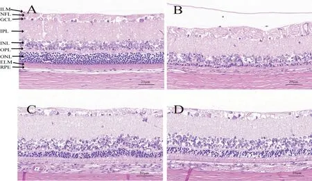
Histopathology of Retina The morphology of the retina of the rats was showed in Figure 4. There were complete overall structure in all groups, but each characteristics were explained below. In the control group, the retina layers were distinct and tight, and the thickness of the retina was about 174.0±7.601 μm. A clear inner limiting membrane (ILM) can be seen; the nerve fibers in the nerve fiber layer (NFL) were arranged well; the nuclei in the ganglion cell layer (GCL) were mostly oval and dense; the inner plexiform layer (IPL) had intact structure; the inner nuclear layer (INL) nuclei were arranged neatly , mostly round and evenly stained; the outer plexiform layer (OPL) had a clear and complete structure; the outer nuclear layer (ONL)nuclei were tightly arranged, oval, darkly stained and about 9-10 layers; the external limiting membrane (ELM) was clear and complete; the cells of the photoreceptor layer of rod and cone were tightly arranged; the retinal pigment epithelium(RPE) was normal in shape, arranged in a short cubic monolayer, and the cytoplasm contained pigment; the blood vessels in the choroid which contained pigment cells were clearly visible, and the boundaries of them could be seen obviously.
在这个供应链平台上,各个节点都将信息公开,让信息透明,使得供应链各个节点都能得到所需的信息,让信息交互更有效率,优化了信息共享的效果,实现共赢。
当下有着巨量的数据,杂乱无章没有经过整理的信息对于企业没有任何意义和作用,所以企业更加迫切需要能够对大数据进行处理和分析的计算机软件以及更多的管理会计人才。这样才能在海量的数据中发掘到对企业有用的部分。面对这样的诉求,企业既可以组织既有员工去学习数据挖掘和分析的知识,也可以招聘具有这方面技能的人员。带动了企业内部的发展,也提升了企业人员的专业水平。
在某日电动汽车充电负荷变化中,如果随机分量较小,b取值可以较大,如果随机分量占比较高,b可以适当减小。这样既考虑到电动汽车充电负荷变化的规律性,又考虑了负荷变化的随机部分。本文的算法流程如图4所示。
在男阴女阳家庭,在女方接受HAART且病毒载量已经控制的情况下可选择体外授精。在男阳女阴家庭选择捐赠精子人工授精可以完全避免HIV传播的风险。如果不接受捐赠精子,也可以在男方进行HAART达到持续病毒抑制后,可考虑在排卵期进行自然受孕。这种情况下夫妻间传染的概率极低[30]。
Ethical Approval This experimental conformed to the standards of the ARVO Statement for the Use of Animals in Ophthalmic and Vision Research. The experiment has passed the animal experiment ethics audit of Fujian Medical University, and the ethics number is LLSLBH-20210625-002.Induction of Animal Model Through LEDs Twenty healthy adult male SD rats, weighing 220±20 g, without eye disease[Shanghai SLAC Laboratory Animal Center, License key:SCXK (Hu) 2017-0005] were randomly divided into 4 groups:the normal control group and three LED exposed groups with low (LED-L), medium (LED-M), and high (LED-H) CRI respectively. Before the experiment, all rats were accustomed to dark for 2d, except for the control group, the rats remained were housed in a breeding cage installed with an LED tube on the top, and the outer periphery of the cage was covered with a light-shielding cloth. Three LED tubes with different CRIs(CRI=10, 83, 95 Ra, 500±50 lx, Sungoing Optoelectronics Technology Co., Ltd., Quanzhou, Fujian Province, China) were installed respectively to irradiate 12h every day, continuing for 4wk, and the remaining 12h was under natural light. During the light exposure, the device was kept ventilated, and the temperature of the light cage was controlled at about 25°C. All rats were fed and drunk freely. The normal control group was fed without any intervention under natural light.
Corneal Fluorescein Sodium Staining The corneal fluorescein sodium staining (CFS) scores were illustrated in Table 3 and Figure 1. The corneal epitheliums of the control group were smooth which were stained only in punctate form after fluorescein sodium staining. Whereas the corneal epitheliums of the LED-M group and the LED-H group were rough and the number of spot stains increased after 2wk, the corneal spot stains of the LED-L group increased significantly and flaky staining appeared meanwhile. After 4wk of LEDs exposure, severe fluorescein plaques appeared in the cornea of the LED-L group and the LED-M group (Figure 2). Compared with the control group (0.20±0.45 points), the differences of the CFS scores in the LED-L group (3.20±0.45 points) and the LED-M group (2.60±0.55 points) were statistically significant(
<0.05) after 2wk. There was no significant difference between the control group and the LED-H group, while there was significant difference between the three LED exposed groups (
<0.05). After 4wk, the corneal scores of the LED-L group (3.80±0.45 points) and LED-M group (3.40±0.55 points)had no statistical difference, while statistically higher than the control group and the LED-H group (
<0.05; Table 3, Figure 1C).
Expression of TNF-α and IL-6 in Lacrimal Gland The result showed that the TNF-α was mainly stained in the cell nucleus. Compared with the control group, the average fluorescence intensity of lacrimal TNF-α in each exposure group had statistically significant differences (
<0.05).Pairwise comparison showed that the average fluorescence intensity of TNF-α in the LED-L group (10.49±2.73) was significantly higher than that of the LED-M group (5.94±2.10)and the LED-H group (4.13±1.37). Meanwhile, the IL-6 was mainly stained in the cytoplasm. The average fluorescence intensity of IL-6 in LED-L group (24.02±5.52) and LED-M group (18.09±5.20) was significantly higher than the control group’s (2.10±0.97;
<0.05). Compared with the light exposed groups, the fluorescence intensity of IL-6 in the LED-H group(3.84±1.80) was significantly lower than that in the LED-L group and LED-M group (
<0.05; Figure 6).
DISCUSSION
The incidence of dry eye has been increasing recently, but its pathogenesis is numerous and still unclear
. The tear film is divided into a mucin layer, an aqueous layer and a lipid layer.And dry eye syndrome is associated with a decrease in tear aqueous production and an abnormality of the lipid, protein,and mucin profiles. Among them, the abnormal decrease in tear aqueous is closely related to dry eye which is secreted by the main lacrimal glands. Studies have shown
that IL-6 and TNF-α are expressed in the ocular surface tissues of rats with exorbital lacrimal gland-excision, indicating the IL-6 and TNF-α play an important role in the pathogenesis of the dry eye.

It’s widely known that the most harmful component of visible light is the blue wavelength (400-500 nm) that can potentially harm eye tissues
, especially the retina
, which can cause inflammation, angiogenesis and so on
. Vicente-Tejedor
removed the blue component of light and found it significantly decreased retinal damage after high intensity exposure. Although the accumulating experimental evidence has showed that exposure to blue light can affect many physiologic functions
, and it can be used to treat circadian and sleep dysfunctions
. But too strong blue light will cause the inhibition of melatonin and affect sleep
. While the impact of LEDs on the lacrimal gland is rarely studied.The international standard CIE218:2016 made the Research Roadmap for Healthful Interior Lighting Applications Toggle navigation that recommended the “healthy lighting” indicator in which mentioned the CRI
. The spectrum emitted by the light determines the light color of the light, and a light with a wider spectral composition is more likely to provide a better color quality
. The current “Hygienic standard for day lighting and artificial lighting for middle and elementary school”issued by the Ministry of Health of the People’s Republic of China
pointed out that the CRI of the classroom lighting source should not be less than 80, especially in professional classrooms such as art, chemistry, and handicrafts, which affects the correct identification of the color of the object prevents the object from displaying its color truly, will cause vision problems such as color blindness and color weakness over time. The white LEDs mainly use blue chip (450-455 nm)and yellow phosphor to generate white light in common
.This method will directly lead to the phenomenon of low CRI and uneven color space distribution, even arise the blue light hazards due to the blue light dominance
. Therefore, we have reason to say that the higher the CRI and the closer to natural light, the better the light color quality may be.


We used three kinds of LEDs with three CRIs from low to high to build a new rat dry eye model. It is observed that the lacrimal gland and retina showed three different degrees of damage after these three kinds of LEDs exposing for 4wk. The immunofluorescence of the inflammatory factor TNF-α and IL-6 in the lacrimal gland showed that with the decrease of CRI, the expression of inflammatory factors increased. The SIt value is the easiest way to reflect the condition of tear aqueous.It’s can be seen that the SIt value of rats in all light exposed groups decreased compared with the control group after 2wk,and it was further reduced until the end of the experiment among which the low CRI (LED-L) group decreased most significantly. These results may suggest that the lacrimal gland exposed to LEDs has undergone morphological changes and inflammation, then affected the normal function of the lacrimal gland, so the aqueous layer was damaged that leaded to a decrease in the SIt.
In this study, a spectrometer (OHSP-350UV, HOPOO Light&Color technology Co., Ltd., Hangzhou, China) was used to measure the spectrum of LEDs (Figure 7). The LED with a CRI of 10 is a pure blue tube, and this spectrum is in the blue wavelength (400-500 nm). The LED with a CRI of 83 has a strong spectral continuity covering the red wavelength(400-700 nm), while its energy is not uniform, of which the energy of the blue wavelength is still the highest. The LED with a CRI of 95 not only has a strong spectral continuity, but a completeness performance that is close to the natural light spectrum. At the same time, it reduces the blue peak value and increases the peak value of green wavelength and red wavelength (500-700 nm), and it has the high saturation and uniformity of various colors closing to the full-spectrum LED.Color rendering is one of the comprehensive effects of the spectrum, especially it has a great relationship with the proportion of the three wavelengths of red, green and blue
.LEDs that have a spectrum closing to the natural spectrum can better replace the ordinary LEDs, which can restore colors,improve visual comfort, and control the peak of blue light and reduce the blue light damage to the eye importantly
.Therefore, the differences of blue light component in these three LEDs spectra may have caused the different levels of inflammatory factors and abnormal morphology of lacrimal glands between the groups, which affected the tears aqueous and caused dry eyes in rats.
All of the exposed groups appeared decrease in retinal thickness,the closer the spectrum is to the full spectrum, the smaller the change in retinal thickness; the higher the blue light component, the more severe the damage to the photoreceptor cell layer. The thickening of the photoreceptor layer of rod and cone in the LED-M group may be a compensatory result of metabolic disorders after this layer being damaged. At the same time, all light exposed groups have increased choroidal thickness and blood vessel density, which may be related to the increase in retinal vascular permeability and compensation after light damage to the blood-retinal barrier, and it remains to be explored later.
The comprehensive effect of the spectrum is affected by a variety of factors, including the color rendering, illuminance,irradiance, correlated color temperature, visible light wavelength and spectral luminous efficiency,
. Therefore, while considering the safety and effectiveness of the light source, it must be considered comprehensively to carry out the optimization of different lighting places. The research in this article not only provides the light selectivity for building the rat dry eye model, but also a certain degree of experimental basis for the optimization of new LEDs and the improvement of lighting standards from the animal experiment level, explains the importance of CRI of LEDs at the level of eye tissue rather than visual.
ACKNOWLEDGEMENTS
Supported by the Natural Science Foundation of Fujian Province (No.2020J01652); the Undergraduate Innovation and Entrepreneurship Training Program of Fujian Medical University (No.YC2003).
Conflicts of Interest: Chen WY, None; Xiao KH, None; Lin R, None; Qiu ZR, None; Chen YM, None; Lin ZQ, None;Ke XB, None; Huang Y, None.
1 Nelson JD, Craig JP, Akpek EK,
. TFOS DEWS II introduction.
2017;15(3):269-275.
2 Huang W, Tourmouzis K, Perry H, Honkanen RA, Rigas B. Animal models of dry eye disease: useful, varied and evolving (Review).
2021;22(6):1394.
3 Lv Y, Chu CC, Liu K, Ru YS, Zhang Y, Lu XX, Gao YC, Zhang CJ,Zhao SZ. A combination of CMC and α-MSH inhibited ROS activated NLRP3 inflammasome in hyperosmolarity stressed HCECs and scopolamine-induced dry eye rats.
2021;11(1):1184.
4 Jung JW, Han SJ, Nam SM, Kim TI, Kim EK, Seo KY. Meibomian gland dysfunction and tear cytokines after cataract surgery according to preoperative meibomian gland status.
2016;44(7):555-562.
5 Pflugfelder SC. Anti-inflammatory therapy of dry eye.
2003;1(1):31-36.
6 Gea M, Schilirò T, Iacomussi P, Degan R, Bonetta S, Gilli G.Cytotoxicity and genotoxicity of light emitted by incandescent,halogen, and LED bulbs on ARPE-19 and BEAS-2B cell lines.
2018;81(19):998-1014.
7 Obana A, Brinkmann R, Gohto Y, Nishimura K. A case of retinal injury by a violet light-emitting diode.
2011;5(3):223-226.
8 Behar-Cohen F, Martinsons C, Viénot F, Zissis G, Barlier-Salsi A,Cesarini JP, Enouf O, Garcia M, Picaud S, Attia D. Light-emitting diodes (LED) for domestic lighting: any risks for the eye?
2011;30(4):239-257.
9 Zhao YN, Ou CL, Yu JK, Zhang YQ, Song HQ, Zhai YP, Tang ZY, Lu SY. Facile synthesis of water-stable multicolor carbonized polymer dots from a single unconjugated glucose for engineering white light-emitting diodes with a high color rendering index.
2021;13(25):30098-30105.
10 Ma N, Li W, Devakumar B, Zhang ZJ, Huang XY. Utilizing energy transfer strategy to produce efficient green luminescence in Ca
LuHf
Al
O
: Ce
, Tb
garnet phosphors for high-quality near-UV-pumped warm-white LEDs.
2021;601:365-377.
11 Fujihara T, Murakami T, Fujita H, Nakamura M, Nakata K.Improvement of corneal barrier function by the P2Y(2) agonist INS365 in a rat dry eye model.
2001;42(1):96-100.
12 Koh S, Watanabe H, Hosohata J, Hori Y, Hibino S, Nishida K, Maeda N, Tano Y. Diagnosing dry eye using a blue-free barrier filter.
2003;136(3):513-519.
13 Tan LL, Morgan P, Cai ZQ, Straughan RA. Prevalence of and risk factors for symptomatic dry eye disease in Singapore.
2015;98(1):45-53.
14 Park B, Jo K, Lee TG, Hyun SW, Kim JS, Kim CS. Polydatin inhibits NLRP3 inflammasome in dry eye disease by attenuating oxidative stress and inhibiting the NF-κB pathway.
2019;11(11):2792.
15 Jaadane I, Villalpando Rodriguez GE,
. Effects of white lightemitting diode (LED) exposure on retinal pigment epithelium
.
2017;21(12):3453-3466.
16 Jaadane I, Villalpando Rodriguez G, Boulenguez P,
. Retinal phototoxicity and the evaluation of the blue light hazard of a new solid-state lighting technology.
2020;10(1):6733.
17 Miralles de Imperial-Ollero JA, Gallego-Ortega A, Norte-Muñoz M,
. Short- and long-term study of the impact of focal blue lightemitting diode-induced phototoxicity in adult albino rats.
2021;22(18):9742.
18 Tao JX, Zhou WC, Zhu XG. Mitochondria as potential targets and initiators of the blue light hazard to the retina.
2019;2019:6435364.
19 Vicente-Tejedor J, Marchena M, Ramírez L, García-Ayuso D, Gómez-Vicente V, Sánchez-Ramos C, de la Villa P, Germain F. Removal of the blue component of light significantly decreases retinal damage after high intensity exposure.
2018;13(3):e0194218.
20 Matynia A, Nguyen E, Sun XP,
. Peripheral sensory neurons expressing melanopsin respond to light.
2016;10:60.
21 Tosini G, Ferguson I, Tsubota K. Effects of blue light on the circadian system and eye physiology.
2016;22:61-72.
22 Lawrenson JG, Hull CC, Downie LE. The effect of blue-light blocking spectacle lenses on visual performance, macular health and the sleepwake cycle: a systematic review of the literature.
2017;37(6):644-654.
23 Wood B, Rea MS, Plitnick B, Figueiro MG. Light level and duration of exposure determine the impact of self-luminous tablets on melatonin suppression.
2013;44(2):237-240.
24 Mou TS, Li JK, Wen XF.
. Proceedings of the 10th China International Semiconductor Lighting Forum; 2013; Beijing China.
25 Hygienic standard for day lighting and Artificial lighting for middle and elementary school: GB 7793-2010. Ministry of Health of the People’s Republic of China; National Standardization Administration of China. 2011.
26 Shen Y, Xie C, Gu YS, Li XY, Tong JP. Illumination from lightemitting diodes (LEDs) disrupts pathological cytokines expression and activates relevant signal pathways in primary human retinal pigment epithelial cells.
2016;145:456-467.
27 Stefani O, Cajochen C. Should we re-think regulations and standards for lighting at workplaces? A practice review on existing lighting recommendations.
2021;12:652161.
28 Bullough J, Bierman A, Rea M. Evaluating the blue-light hazard from solid state lighting.
2019;25(2):311-320.
29 Zhu Y, Valter K, Stone J. Environmental damage to the retina and preconditioning: contrasting effects of light and hyperoxic stress.
2010;51(9):4821-4830.
30 Perikala M, Bhardwaj A. Excellent color rendering index single system white light emitting carbon dots for next generation lighting devices.
2021;11(1):11594.
31 Huang Z, Chen W, Liu Q, Wang Y, Pointer MR, Liu Y, Liang JX.Towards an optimum colour preference metric for white light sources:a comprehensive investigation based on empirical data.
2021;29(5):6302-6319.
32 Touitou Y, Point S. Effects and mechanisms of action of lightemitting diodes on the human retina and internal clock.
2020;190:109942.
猜你喜欢
珠江水运(2022年7期)2022-05-12
中国典型病例大全(2022年13期)2022-05-10
健康体检与管理(2022年2期)2022-04-15
中国医药导报(2019年16期)2019-09-07
中国实用医药(2018年12期)2018-05-03
科技创新与应用(2017年15期)2017-05-31
价值工程(2017年9期)2017-04-18
现代企业文化·理论版(2016年20期)2016-12-20
建材发展导向(2014年5期)2014-10-20
城市建设理论研究(2011年23期)2011-12-20
 International Journal of Ophthalmology2022年7期
- International Journal of Ophthalmology的其它文章
- Impact of OCT scan-patterns in identifying morphological features of lamellar macular holes and macular pseudoholes
- Virtual reality training improves accommodative facility and accommodative range
- Short-term effect of 0.01% atropine sulphate eye gel on myopia progression in children
- Reduced choroidal peripapillary capillaries in thyroidassociated ophthalmopathy with early stage of dysthyroid optic neuropathy
- Incidence of ocular manifestations in patients with graft versus host disease after allogeneic stem cell transplant in Riyadh, Saudi Arabia
- Clinical features, surgical outcomes and genetic analysis of ectodermal dysplasia with ocular diseases