
Congenital retinal macrovessel with intracranial venous malformation in a pediatric patient: a case report
2022-07-30 10:03HyeonJinParkSookHyunYoonSookYoungKimDonghunLee
Dear Editor,
Congenital retinal macrovessel (CRM) is defined as an abnormal large retinal vessel that encircles the foveal avascular zone (FAZ)
. Since the first description of a large aberrant vessel crossing the macula by Mauthner
in 1868,there have been several case reports about CRM
. And recently, Pichi
reported clinical manifestations and multimodal retinal imaging of 49 eyes with CRM. In their study, they had identified an association between macrovessel in the retina and venous anomalies of the brain and they emphasized the importance of systemic workups including brain magnetic resonance imaging (MRI). Meanwhile,previous reported cases were almost adults and abnormal macular vessel in pediatric patient was rarely reported
.Souissi
reported a case of 15-year-old girl with congenital hamartoma associated with CRM that resulted in the mild visual impairment. However, systemic evaluation including brain imaging was not performed.
Here, we report a case of CRM in a pediatric patient aged 4y with cerebrovascular analysis through brain MRI with magnetic resonance angiography (MRA) and retinal vessel analysis using optical coherence tomographic angiography(OCTA).
Ethical Approval The study was conducted in accordance with the Declaration of Helsinki. Informed consent for publication was obtained from the family of the patient in this case report.
Case Report A girl aged 4y presented to the Ophthalmic Department with the complaint of frequent eye blinking for a month. The patient had no other history of ocular trauma and no known ocular or systemic disease. Her mother also had no medical history during the perinatal period. The visual acuity was 20/40 in both eyes, and there was no afferent pupillary defect. On slit-lamp examination, mild punctate epithelial erosions on her left eye were observed. For revealing her refractive errors, cycloplegic refraction was performed and+3.00 diopters of hyperopia with +1.00 diopters of astigmatism in both eyes was noted.
但回国的道路并不顺利。刘万传讲述了归国途中遇到的炮击:“我们以步行为主,有一段是坐汽车,部队交替撤退,工兵最后布雷。在撤军途中,越南的炮火打了过来,他们重炮很猛的,很多都是1 0 0毫米口径以上的,我们遇到炮击后,就赶快找掩体躲避起来。很多新补充的新兵不知道怎么躲,没有经验,没能回来。”
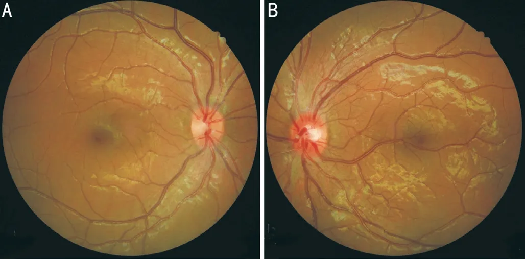
There were no remarkable findings on right fundus examination (Figure 1A); however, the left fundus revealed multiple branches of retinal vessels and a large aberrant central retinal vein originating from the inferotemporal disc cup that transverses the macula (Figure 1B). On OCTA images(AngioVue; Optovue, Fermont, CA, USA), the aberrant macrovessel crossed the foveal area beyond the horizontal raphe (Figure 2A), and thin retinal vessels branching from the macrovessel ran through the foveal avascular zone (FAZ;Figure 2B). Moreover, spectral domain OCTA revealed an irregular contour of the inner retinal layers owing to multiple superficial retinal vessels (Figure 2C). There was no exudate,hemorrhage, cyst, or leakage on the retina.
The current report is meaningful since the patient was the youngest ever, compared to previously reported cases,and intracranial vascular malformation was also noted.Morphologically, CRM can be venous, arterial
, and sometimes arteriovenous communications
. In Pichi
’s
reports with a large series of eyes with CRM, early phase frames of fluorescein angiography (FAG) further confirmed the venous nature of the CRM. However, in this case, FAG could not be performed since the patient was young. Instead, OCTA was performed, and as previously described, microvascular capillary abnormalities around the CRM were evident in the deep capillary plexus image. Moreover, they described that 24% of enrolled patients with CRM had potential cerebral associations. Likewise, this pediatric patient also had asymptomatic cerebrovascular abnormalities, dilated superior ophthalmic vein, and fenestration of the basilar artery.
Since there were no other symptoms associated with CRM on the left eye, the patient was managed conservatively using glasses to correct hyperopic compound astigmatism. Use of artificial tears for treating corneal erosion was recommended.After 6-month of observation while wearing hyperopic glasses,her best-corrected visual acuity improved to normal range without any evidence of amblyopia. Her eye blinking also improved. On fundus examinations, the CRM remained stable,and complications associated with macrovessels were not observed.
For excluding other cerebrovascular malformations, MRI with MRA of the brain was performed and there was no significant abnormality in the brain parenchyma. However, in the axial three-dimensional time-of-flight image of the MRI, a slightly dilated left superior ophthalmic vein was noted (Figure 3A).Additionally, MRA revealed a small fenestration of the proximal basilar artery (Figure 3B).
Previous studies on retinal macrovessels are mostly in the form of case reports
; therefore, the accurate prevalence of CRM is unclear; however, Hayasaka
reported that unusual retinal vessel patterns were found in 21 of 3506 eyes in their retrospective fundus photographs analysis. Meanwhile,among previous case reports of CRM, pediatric cases have only rarely been reported
. de Crecchio
described a patient aged 5y with an anomalous retinal vein crossing the macular and touching the foveal area. Visual impairment occurred by the mere presence of a foveal lesion. However,there is a limitation that a systemic examination including brain imaging was not performed to reveal any other lesions related to ophthalmic findings.

我们恰恰就是想通过这些摄影艺术作品的多元性和相通性,展现出不同国家、地区、文化和民族的摄影发展趋势,从而充分体现出多元与融合的摄影理念,以此增进国际间的了解,加强中国与其他国家、地区的对话与合作,用发展的眼光探讨人类对当下与未来的展望。

“希望来吧”作为团省委关爱流动和留守儿童的主阵地,既给予了流动和留守儿童关爱与帮助,又凝聚了各级团组织、青年志愿者以及社会爱心人士的志愿力量,同时,还加强了基层团组织的建设。
There have been several hypotheses regarding the presence of aberrant retinal vessels in the FAZ. Embryologically, the development of FAZ is debatable. Some authors propose that the FAZ develops by “apoptotic remodeling” after 36wk of gestation
, while others report that retinal vasculature developed towards the fovea and the foveal region was normally avascular during all gestational periods
. CRM refers to the presence of aberrant vasculature where capillaries are usually absent, and Bhatia
reported that vascular proliferation may reach the foveola if foveal hypoxia occurs and CRM develops. Meanwhile, Ashton
explained that CRM develops around 15-16wk of gestation when differentiation of retinal arteries and veins occurs. Moreover, the origin of CRM is probably related to hemodynamic variation. In our patient, there was no previous history of ischemic risk, such as maternal gestational diabetes; therefore, so it is unlikely that any systemic disease that could cause foveal hypoxia and development of CRM. Using Ashton’s
embryologic concept, it is possible that large interindividual variation in cerebrovascular and retinal vascular anatomy occurred during the gestational period. Most studies have found that CRMs are asymptomatic and visual acuity is preserved
. Likewise, our case did not have any discomfort associated with CRM. Her mild visual impairment at the initial visit in the ophthalmologic clinic had improved after wearing glasses; therefore, it was assumed to be owing to her refractive errors rather than the macular lesion. However, visual impairment may be caused by preretinal hemorrhage or by the mere presence of the macrovessel in the foveal area
. de Crecchio
described that 5 of 13 cases complained of visual impairment caused by the CRM in their study. Vitreous hemorrhage
,macroaneurysms
retinal ischemia
, or macular edema
originating from the CRM have also been reported. Therefore,even if there is no visual discomfort in patients with CRM,regular follow-up in an ophthalmology clinic should be considered to check changes in the macular lesion.
Analysis of impact of rail transit construction sites on surrounding road traffic and traffic organization optimazation
CRM can be detected in asymptomatic pediatric patients. In addition to ophthalmic examinations, careful neuroimaging should be considered to reveal other cerebrovascular malformations. For pediatric patients, regular follow-up is necessary considering the complications that are associated with CRM, such as aneurysms, retinal hemorrhage, or amblyopia.
ACKNOWLEDGEMENTS
Conflicts of Interest: Park HJ, None; Yoon SH, None; Kim SY, None; Lee D, None.
1 Pichi F, Freund KB, Ciardella A, Morara M, Abboud EB, Ghazi N, Dackiw C, Choudhry N, Souza EC, Cunha LP, Arevalo JF, Liu TYA, Wenick A,He LM, Villarreal G Jr, Neri P, Sarraf D. Congenital retinal macrovessel and the association of retinal venous malformations with venous malformations of the brain.
2018;136(4):372-379.
2 Mauthner L.
. Vienna, Austria:Tendler,1868.
3 Preziosa C, Milani P, Ciasca P, Bergamini F, Staurenghi G, Pellegrini M. Optical coherence tomography angiography findings in a case of congenital retinal macrovessel with anomalous retinal anastomosis associated with contralateral myelinated nerve fibers and retinal vascular abnormalities.
2021;15(5):605-610.
4 Jiang X, Zheng C, Du F, Ai S. Multimodal imaging of aberrant macular microvessel crossing the foveal avascular zone in two young adults.
2020;20(1):207.
5 Tripathy K, Bypareddy R, Chawla R. Congenital retinal macrovessel may be associated with unilateral foveal hypoplasia/small foveal avascular zone.
2019;54(1):139.
6 Hasegawa T, Ogata N. Retinal deep capillary ischemia associated with an occluded congenital retinal macrovessel.
2017;11(3):277-280.
7 Souissi K, El Afrit MA, Kraiem A. Congenital retinal arterial macrovessel and congenital hamartoma of the retinal pigment epithelium.
2006;43(3):181-182.
8 de Crecchio G, Alfieri MC, Cennamo G, Forte R. Congenital macular macrovessels.
2006;244(9):1183-1187.
9 Hayasaka S, Katsube T, Ugomori S, Setogawa T. Abnormally distributed branches of the retinal vessels, enlarged macular arteries and long cilioretinal arteries.
1990;200(4):194-197.
10 Brown GC, Donoso LA, Magargal LE,
. Congenital retinal macrovessels.
1982;100(9):1430-1436.
11 Bhatia HK, Sharma S, Laxminarayana P. Congenital retinal macrovessel with normal visual acuity: a case report.
2015;2(2):017.
12 Jager RD, Timothy NH, Coney JM, Katalinic P, Cavicchi RW, Strong J, Cavallerano JD, Aiello LP. Congenital retinal macrovessel.
2005;25(4):538-540.
13 Chalam KV, Gupta SK, Vinjamaram S, Shah VA. Clinicopathologic reports, case reports, and small case series: congenital anomalous retinal artery associated with a leaking macroaneurysm.
2003;121(3):409-410.
14 Mintz-Hittner HA, Knight-Nanan DM, Satriano DR, Kretzer FL. A small foveal avascular zone may be an historic mark of prematurity.
1999;106(7):1409-1413.
15 Provis JM, Hendrickson AE. The foveal avascular region of developing human retina.
2008;126(4):507-511.
16 Ashton N. The mode of development of the retinal vessels in man. The William MacKenzie centenary symposium on the ocular circulation in health and disease: Mosby, 1969:7-17.
17 Strampe MR, Wirostko WJ, Carroll J. A case of congenital retinal macrovessel in an otherwise normal eye.
2017;8:18-21.
18 Goel N, Kumar V, Ghosh B. Congenital retinal macrovessel associated with vitreous hemorrhage.
2017;21(1):83-85.
19 Sebrow DB, Cunha de Souza E, Belúcio Neto J,
. Macroaneurysms associated with congenital retinal macrovessels.
2020;14(1):61-65.
20 Medina-Tapia A, Molina-Sócola FE, Llerena-Manzorro L, López-Herrero F, Castilla-Martino M, Martínez-Borrego A, Sánchez-Vicente JL. Congenital retinal macrovessel associated with retinal peripheral telangiectasia and retinal ischaemia.
2017;92(7):338-342.
21 Zheng F, Lai K, Yin H, He J, Xu Y, Ye P. Case report: self-resolved macular edema secondary to congenital retinal macrovessels.
2022;8:771007.
22 Ipek SC, Kavukcu S, Men S, Saatci AO. Multimodal imaging features of a spontaneously resolved unilateral congenital macrovessel-related macular edema in a 13-year-old boy.
2020;10:Doc40.
猜你喜欢
理财·市场版(2022年5期)2022-05-30
经济研究导刊(2020年6期)2020-04-10
新青年(2020年1期)2020-03-07
艺术研究(2016年4期)2017-01-16
未来英才(2016年13期)2017-01-13
都市家教·下半月(2016年11期)2016-12-29
考试周刊(2016年95期)2016-12-21
青春岁月(2016年21期)2016-12-20
文理导航(2016年33期)2016-12-19
青年与社会(2016年31期)2016-11-28
 International Journal of Ophthalmology2022年7期
- International Journal of Ophthalmology的其它文章
- Impact of OCT scan-patterns in identifying morphological features of lamellar macular holes and macular pseudoholes
- Virtual reality training improves accommodative facility and accommodative range
- Short-term effect of 0.01% atropine sulphate eye gel on myopia progression in children
- Reduced choroidal peripapillary capillaries in thyroidassociated ophthalmopathy with early stage of dysthyroid optic neuropathy
- Incidence of ocular manifestations in patients with graft versus host disease after allogeneic stem cell transplant in Riyadh, Saudi Arabia
- Clinical features, surgical outcomes and genetic analysis of ectodermal dysplasia with ocular diseases