
Occult suprachoroidal foreign bodies: a 3-case report
2022-07-30 10:03HuPingSongRongLeZhouZhaoLiangZhuTaoChenJianZhouWang
Dear Editor,
Intraocular foreign bodies (IOFB) account for 18% to 41%of open globe injuries (OGI), which can cause severe vision loss and bring a heavy economic burden to the patients and society
. Most of the IOFB (58%-88%) are located in the ocular posterior segment
. Foreign bodies (FB) in the suprachoroid are rare
. From January 1, 2015 to April 30,2020, there were 2110 cases of OGI in Ocular Trauma Center of Xi’an Fourth Hospital, and 3 cases of suprachoroidal FB were detected among 409 cases of IOFB. Now we reported these cases.
Case 1 A 35-year-old man suffered from an ocular injury of the right eye when striking metal, and he was referred to the Emergency Department (Table 1). The uncorrected visual acuity (VA) was hand motion (HM) in the right eye. There was a 5 mm length scleral penetrating wound, which was 8 mm posterior to the limbus, nasally to the inferior rectus muscle, with vitreous prolapse. The lens was clear, and the intraocular pressure (IOP) was normal. Owing to vitreous hemorrhage (VH), fundus examination was not possible with a poor view. Computed tomography (CT) scanning of orbits demonstrated a hyperdense shadow of the metallic fragment in the supratemporal wall of the eye (Figure 1A and 1B). The surgery was performed under general anesthesia on the day of admission. The scleral wound was sutured and vitrectomy was performed. After VH was removed, a retinochoroidal break corresponding to the site of the scleral wound was found, and the retina detached. Another retinal break was noted 2 optic disc diameters (DD) superior temporal to the fovea, the retina detached, where the choroidal break was not identified, and no FB could be seen in the vitreous cavity or subretinal space.According to the clinical manifestation and CT data, the FB was considered to be within the eyeball wall, but not be an IOFB. And then, argon laser retinopexy was applied around the retinal break. Silicone oil was used as intravitreal tamponade.Two weeks later, the patient presented for follow-up. Fundus examination revealed an oval choroidal elevation of 2-3 DD,which was adjacent to the superior temporal retinal break. The tissue fibrosis of retinal and choroidal breaks at the sites of entrance and exit of FB was noticed (Figure 1C). The orbital CT scanning confirmed nearly identical location of the IOFB to that of preoperational. Then suprachoroidal FB was diagnosed.After that, an operation of FB removal was performed. During the operation, the retina and choroid overlying the IOFB were cauterized. The retinochoroidotomy was made at the site of choroidal elevation. The IOFB, measuring 5×3×2 mm
, was finally removed from the vitreous cavity through the enlarged scleral incision, by an intraocular magnet (Figure 1D and 1E).Following removal of the FB, the inner surface of the scleral wall was exposed apparently. Endolaser was applied around the incision of retinochoroidotomy, and silicone oil was used as tamponade. Three months post-operation, silicone oil was removed. The uncorrected VA of the right eye improved to 20/200 without recurrence of retinal detachment (RD).
如表1所示,原序列的ADF值大于5%临界值,不能拒绝原假设,表明原序列数据不平稳;而原序列一阶差分的ADF值小于5%临界值,可以拒绝原假设,故可认为是平稳序列,即ln(crmb)、ln(ix)、ln(ex)是一阶单整序列。
1.2.2 实验组 分康复评定、治疗技术、病例分析三个考站,所有考生必须按顺序完成三站内容的考核,其中评定与治疗两考站的内容由主考老师于所有内容中分别抽取3项,共6项,所有考生考核内容相同,评分标准一致,具体内容如下:
Case 2 A 36-year-old male presented with vision loss in his left eye for 6h. His left eye was hit by something while hammering metal. He was healthy in the past. The VA of the left eye was light perception (Table 1). By slit-lamp biomicroscope, nasal subconjunctival hemorrhage, and conjunctival laceration, a 10 mm irregular transverse scleral laceration at 9 o’clock, 2 mm posterior to the limbus, with vitreous prolapse, and corneal edema were revealed. The anterior chamber was shallow, with little hyphema, and the lens was mildly opacified, and the fundus could not be seen clearly because of VH. A metal IOFB of 4×6 mm
in the vitreous cavity posteriorly and inferiorly in the left eye was detected by CT scanning. On the day of admission, the globe was repaired, with vitreous injection of vancomycin, and systemic antibiotics were prescribed. On the second day, the pars plana vitrectomy (PPV) was performed. During the surgery, we found a retinochoroidal break inferotemporal to the fovea, with large subretinal hemorrhage, and RD. A choroidal elevation of 2 DD was noted about 2 DD inferior to the retinochoroidal break. No obvious IOFB was found in the vitreous cavity, after the removal of VH, then tamponaded with silicone oil.

IOFB is common. Posterior segment FB is more likely to cause comorbidities, such as traumatic cataract, VH, and RD,associated with marked loss of vision
. Nearly 52.38% of FBs were located in the vitreous, 33.33% were in the retina,and 14.28% were in perforating
. While suprachoroidal FB is rarely reported.
对9度地震烈度下钢管塔沿塔身高度的位移响应进行研究,分别在4类场地下沿0°方向输入地震波,分别计算5.17,15.5,20.5,26.6,39.0,50.5,71.5,92.0,95.5,101 m塔身高度下的位移响应,得到的钢管塔沿塔身高度的位移响应曲线如图4所示.
CT scanning is one of 3 main imaging tools used in the diagnosis of OFB
. Previous studies showed that the sensitivity of CT scanning in the diagnosis of OFB was 61.7%,the specificity was 100%, and the accuracy in locating FB was 94.7%. But in the condition of the FB were metal foreign objects, with a diameter of 4 to 5 mm, and adjacent to the eyeball wall, they were easy to be misdiagnosed as eyeball wall instead of an IOFB. It is likely due to the overlapping of FB shadows and the eye ring
. In our cases, the FB consistent with all of the terms above mentioned, meanwhile, during the first operation, the FB was not found intraocular, so we misdiagnosed it as an orbital or eyeball wall FB.
The suprachoroidal FB was rarely referred to, maybe owing to its low incidence, although ocular foreign bodies (OFBs)can be classified into IOFB and extraocular FB depending on whether they are within the eyeball or outside it
, or it can be described as global FB (anterior chamber, iris, lens, vitreous,choroid or retina), adnexal FB (in orbit, lids, conjunctiva and lacrimal apparatus) and mixed
. From January 1, 2015 to April 30, 2020, there were only 3 cases of suprachoroidal FB, in Xi’an Fourth Hospital, a tertiary hospital in Xi’an city,which has an independent ocular trauma center. It accounted for 1.42% of the 2110 consecutive OGI, and 7.33% of 409 IOFB. Two of the three patients suffered from ocular trauma while striking mental, which is the most frequent ocular trauma mechanism in China
.
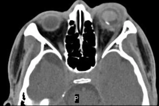
Case 3 A 30-year-old male worker presented with vision loss in his left eye for 3h after hammering concrete in a construction site(Table 1). His VA of the left eye was HM before the eye. There was a scleral wound of 5 mm at 7 o’clock, 5 mm posterior to the limbus, with vitreous prolapse. The lens was opacified. The fundus couldn’t be seen clearly because of VH. The orbital CT displayed that there was a metallic FB (6×3 mm
) in the left eye supratemporal, with the CT value of 2963 Hu (Figure 3A). The surgery was performed on the first day of admission.Multiple 7-0 Vicryl interrupted sutures were used to close the scleral laceration. PPV was performed. A retinochoroidal break corresponding to the scleral wound was obvious, while another retinal break was found, about 2 DD supratemporal to the fovea, with localized retinal edema, hemorrhage, and mild RD,but no choroidal break was seen in the corresponding site. An oval choroidal elevation was noticed, which was about 4 DD supratemporal to the retinal break. No obvious FB was found in the vitreous cavity or subretinal space. The operation was stopped. Silicone oil was used as tamponade.
One day after the vitrectomy, a second orbital CT scanning was made, a similar shape of IOFB at the similar location was identified (Figure 2). After communication with the patient and his wife, they agreed to take the second surgery to remove the FB 1wk later. During the second surgery, a metallic FB was revealed finally in the site of choroidal elevation following retinochoroidotomy, and it hid in the suprachoroidal cavity of the corresponding site.


On the first day post-surgery, the second orbital CT scanning was taken and detected the metal FB with nearly the same shape and location as that before (Figure 3B). No obvious FB was found by fundus examination (Figure 3C). By optical coherence tomography, especially between the macular and superior arcade, no FB was detected because of the limits of detection depth (Figure 3D). It was doubted that the FB be hidden under the elevation of the choroid, so the second surgery was performed, 7d after the first operation. After the retinochoroidotomy was performed at the site of elevation of choroidal, a metal FB (3×5×2 mm
) was found. It was removed through the enlarged incision of the cornea successfully with an intraocular magnet. The diathermy and endolaser were done and tamponaded with silicone oil. One month post-operation,the VA was counting fingers at 40 cm, with normal IOP.
The first case was reported by Hashim
. That patient had one self-sealed scleral penetrating wound, 3 mm posterior to the limbus, with a retinochoroidal break exposing the bare sclera temporally just posterior to the equator. An oval choroidal elevation of 1 DD was noted 2 DD temporal to the fovea, with a small line of choroidal elevation continuous from the retinochoroidal break. The retinochoroidal break and choroidal elevation were identified apparently. The FB was removed successfully during the first operation 3wk later. Different from Hashim
’s
case, in cases 1 and 3,the scleral entrance wound was evident, while there was no obvious exposed choroidal break in the exit site of the eyeball,but only a retinal break. Therefore, during the first operation,the IOFB was not found or removed successfully. They can be described as occult suprachoroidal FB, similar to occult scleral rupture. It was speculated that the FB penetrated the eyeball wall, and created an obvious retinal break, then it was deflected by the rigid scleral wall, and so traversed in the suprachoroidal space, and finally resided in the suprachoroidal space to form a choroidal elevation.
岗位工作内容是职业标准的一个组成部分,国家职业标准具体列举了职业的完整工作内容。国际商法课程的项目不应该来源于传统教材的目录单元或教师的主观构想,而应来源于职业标准中工作内容,它是国际商事活动中具有相对独立性的一系列典型具体工作。
Removal of this posterior suprachoroidal FB required retinochoroidotomy. Any form of hemorrhage at this site is potentially devastating to the central vision. In the case of Hashim
, hypotensive anesthesia was used as a means of providing excellent hemostasis during the removal of the suprachoroidal FB. In our cases, we only used general anesthesia, no hypotensive anesthesia was emphasized, we did not encounter any form of strong hemorrhage, so we presumed that hypotensive anesthesia may not be imperative.
In conclusion, the characteristics of the above 3 cases includes:a full-thickness break of the retina caused by a high-velocity projectile passing adjacent to, but not perforating the globe;CT scanning showed adjacent to eyeball wall FB and the elevation of the choroid. Under these conditions, the occult suprachoroidal FB should be considered as a diagnosis.
ACKNOWLEDGEMENTS
Thanks for the valuable advice and guidance of Dr. Jing-Bo Wang.
Conflicts of Interest: Song HP, None; Zhou RL, None; Zhu ZL, None; Chen T, None; Wang JZ, None.
1 Loporchio D, Mukkamala L, Gorukanti K, Zarbin M, Langer P,Bhagat N. Intraocular foreign bodies: a review.
2016;61(5):582-596.
2 Ma J, Wang Y, Zhang L, Chen M, Ai J, Fang XY. Clinical characteristics and prognostic factors of posterior segment intraocular foreign body in a tertiary hospital.
2019;19(1):17.
3 Hashim H, Lim KS, Choong YY, Nor NM. Hypotensive anesthesia in the management of a posterior suprachoroidal foreign body.
2005;25(1):87-89.
4 Mukkamala LK, Soni N, Zarbin MA, Langer PD, Bhagat N. Posterior segment intraocular foreign bodies: a 10-year review.
2017;1(4):272-277.
5 Nicoară SD, Irimescu I, Călinici T, Cristian C. Intraocular foreign bodies extracted by pars plana vitrectomy: clinical characteristics,management, outcomes and prognostic factors.
2015;15:151.
6 Kuhn F, Morris R, Witherspoon CD. Birmingham Eye Trauma Terminology (BETT): terminology and classification of mechanical eye injuries.
2002;15(2):139-143,v.
7 Shukla B. New classification of ocular foreign bodies.
2016;19(6):319-321.
8 Wang WP, Zhou YL, Zeng J, Shi M, Chen BH. Epidemiology and clinical characteristics of patients hospitalized for ocular trauma in South-Central China.
2017;95(6):e503-e510.
猜你喜欢
中国质量与标准导报(2022年2期)2022-06-09
孩子(2020年8期)2020-08-13
诗潮(2019年1期)2019-01-25
启迪与智慧·下旬刊(2019年9期)2019-01-19
读者(2018年8期)2018-04-03
共产党员·下(2017年1期)2017-02-09
科技与企业(2015年15期)2015-10-21
安徽文学(2015年8期)2015-09-10
新高考·高二数学(2014年7期)2014-09-18
环球时报(2009-11-25)2009-11-25
 International Journal of Ophthalmology2022年7期
- International Journal of Ophthalmology的其它文章
- Impact of OCT scan-patterns in identifying morphological features of lamellar macular holes and macular pseudoholes
- Virtual reality training improves accommodative facility and accommodative range
- Short-term effect of 0.01% atropine sulphate eye gel on myopia progression in children
- Reduced choroidal peripapillary capillaries in thyroidassociated ophthalmopathy with early stage of dysthyroid optic neuropathy
- Incidence of ocular manifestations in patients with graft versus host disease after allogeneic stem cell transplant in Riyadh, Saudi Arabia
- Clinical features, surgical outcomes and genetic analysis of ectodermal dysplasia with ocular diseases