磁共振氨基质子转移成像技术原理和应用
2016-04-17 04:56吴仁华
磁共振成像 2016年4期
吴仁华
磁共振氨基质子转移成像技术原理和应用
吴仁华*

本文概述了氨基质子转移(amide proton transfer, APT)技术与化学交换饱和转移(chem ical exchange saturation transfer, CEST)技术的关系、APT技术的原理、影响APT技术的诸多因素,特别是在体APT成像需研究核奥氏效应(nuc lear Overhauser effect, NOE)的重要性。阐述了APT技术的应用,主要有pH成像、温度成像、APT加权像在各器官组织的临床应用。
氨基质子转移;化学交换饱和转移;核奥氏效应;酸碱度成像;温度成像
Received 17 Feb 2016, Accepted 3 Apr 2016
ACKNOW LEDGM ENTSThis work was part of National Natural Science Foundation of China (No. 81471730).
磁共振氨基质子转移(am ide proton transfer, APT)成像[1-4]是化学交换饱和转移(chem ical exchange saturation transfer, CEST)技术[5-6]的一种,CEST技术与磁化传递技术[7-8]密切相关,从某种意义来说,CEST技术是特异性预饱和的磁化传递技术,而临床上应用的磁化传递成像是非特异性饱和的磁化传递技术。尽管CEST成像目前还有许多问题尚待解决,特别是在体CEST成像主要会有直接水饱和(溢出效应)、大分子交换池(磁化传递)和核奥氏效应(nuclear overhauser effect, NOE)等的影响[9-10],由于CEST成像具有高空间和时间分辨率的优点[11-13],可检测蛋白和代谢物中的可交换质子,用于无创伤的酸碱度(pH值)成像[1, 4]、内源性蛋白分子成像[14-15]、代谢物成像[16-17]、细胞标记[18-19]、报告基因的研究[20]等,目前是磁共振成像研究的热点。
CEST技术中的APT成像在模型、动物和人体层面上有过非常多的研究[9, 11, 14, 16, 21],学术界有过广泛的讨论,目前认为在优化技术的前提下,在人体层面上可以获得APT加权像[22],APT加权像具有重要的临床应用价值,特别是应用于脑肿瘤的诊断和鉴别诊断[3, 23-25]、脑卒中的早期诊断[26-27]、神经变性疾病的定性[28-29]、肝糖原检测[30]等,本文主要综述APT成像的技术原理和临床应用价值。
1 氨基质子转移成像技术原理
1.1 氨基质子的预饱和频率
APT成像的技术原理也基于CEST技术的原理,只是APT成像的预饱和脉冲频率是特异性饱和氨基质子。Forsen等[31]在1963年利用双共振方法开创性地研究了快速化学反应现象。Guivel-Scharen等[32]在1998年研究小分子溶液的磁化传递现象时最早观察到靠近水共振频率Z谱的不对称性,为了与既往的磁化传递名词相区分,将这一磁共振现象命名为 CEST。这些学者发现水的氢质子(4.7 ppm)与内源的游离蛋白质和多肽上的酰胺基(-NH)质子(8.3 ppm)存在一个快速的化学交换(氢交换)的过程,而且此过程与环境的pH值相互依赖,这一磁共振现象随之被命名为氨基质子转移[13, 33-35]。
1.2 CEST技术原理的二池、三池模型
对CEST技术原理最经典的解释是二池模型(图1)[13, 27, 36],假设人体内环境中有如下两池(A、B池),A池代表体内水质子信号,B池代表体内某游离大分子的某个(或几个)质子信号,在主磁场中(B0)二者会有着不同的共振进动频率,通过施加选择性的脉冲(预饱和脉冲B1)饱和B池信号,在适当的条件下(适宜的温度、pH范围),这些质子会和周围的水质子发生化学交换,进而将部分饱和传递到A池,通过特定磁共振技术采集到的A池信号可以反映出该CEST效应的强弱效果[13, 33-35]。
也有学者提出磁共振CEST技术原理的三池模型及其它模型,所谓三池模型即是将组织中的质子分为三个池。第一个池为自由水池,为磁共振成像提供大量信号。第二个池为结合池,包含与蛋白质、其他大分子或者细胞膜束缚的氢质子,主要是人体中的半固态组织,这些氢质子的信号在常规磁共振成像技术中是无法被检测的。第三池为特定分子池,系游离大分子(某些内源游离大分子或CEST对比剂)上的氢质子(包括某些羟基、胺基、酰胺基及亚胺基等)为可交换的质子。CEST技术主要研究的是第三池与自由水池的交换效应,但结合池的影响不可忽视[27, 33-37]。
1.3 外源性化合物的研究
APT成像主要依赖内源性的氨基质子,当应用某些外源性化合物时,如镧系螯合物[38]、CT扫描的对比剂[39]等,氨基质子的交换会发生变化,这对于酸碱度的研究有一定意义。
1.4 核奥氏效应(NOE)
应用化合物模型对APT成像进行研究时,可以得到非常规整的CEST谱(Z谱)和效果非常好的APT图,但在体研究时,特别是人体研究时,Z谱不可能规整,其中主要是受到NOE的影响,因而目前认为在人体层面上获得的是APT加权像[22]。NOE的定义可以这样表述:当某一自旋的核磁共振(nuclear magnetic resonance, NMR)吸收得到饱和时,另一自旋的NMR吸收强度积分值的改变[40]。在分析Z谱时,认为氨基质子对侧的谱线为NOE效应[41-43]。要深入研究APT成像,必须研究NOE的影响及其NOE成像。
2 氨基质子转移成像技术的应用
2.1 在体器官组织的pH测定
APT成像技术已有很广泛的应用,其中一个主要方向是在体器官组织的pH测定[1, 4, 44-46]。相对于磁共振谱方法的pH成像,APT成像方法具有高空间和时间分辨率。正常的人体机能有赖于正常的稳定的内环境,组织pH的改变是提示许多病理变化的指标。肿瘤细胞内pH(pH i)及细胞外pH (pHe)存在梯度差。与正常组织相比,pHe通常较低,而pHi可以较高或基本无变化。因此,通过检测pH的变化可评估肿瘤病人的预后及治疗反应[45]。APT成像的pH研究大部分在3 T以上场强磁共振机器上进行,笔者研究在临床1.5 T机器上也可以实现pH的检测[4](图2)。
对于pH的测定,目前的研究方向是全面考虑影响pH测定的各种因素[47],进而精准无损伤地在体量化器官组织的pH值[48],不仅量化细胞内pH (pHi),结合外源性对比剂[39, 49],也量化细胞外pH (pHe)值,有助于良性肿瘤与恶性肿瘤的鉴别,也有助于肿瘤异质性的判定,观察一些疾病的治疗效果。
2.2 在体器官组织的温度测定
结合外源性对比剂[50],应用氨基质子转移技术可以在体测定pH值的同时,测定组织的温度差异。结合水交换谱(w ater exchange spectrum, WEX),不应用外源性对比剂也可以获得相似的pH和温度信息,该技术也有在体应用的潜力[51],无损伤的在体器官组织磁共振温度测定相对于红外线温度测定,其内部结构更清楚,有望应用于肿瘤射频治疗的温度监测。
2.3 在各器官组织的应用
2.3.1 在脑的应用
APT技术早期应用于脑梗塞的研究,认为APT成像比弥散加权像在显示梗死组织方面更有优势[1],在判断脑梗死半暗带方面也有优势[52],能更精确判断脑梗死半暗带的范围,这对于是否选择溶栓治疗非常有帮助。APT成像在脑梗死方面的研究一直持续至今[52-55],在超急性期脑卒中的动物中,可简便地区分开出血性脑中和缺血性脑卒中[55]。APT技术同时也较多地应用于脑肿瘤方面的研究[3, 24, 56],研究认为APT技术有助于脑肿瘤的诊断、鉴别诊断和治疗,应用APT技术显示胶质瘤的内部结构,可以与应用对比剂时才能获取的信息相媲美,可应用于转移瘤和高级别神经上皮肿瘤的鉴别诊断。在神经变性疾病方面,APT技术有助于神经变性疾病的早期诊断,可作为帕金森病临床诊断和病情监测的有效工具,为帕金森病的病理生理研究提供重要的信息[29]。
2.3.2 在肾脏的应用
结合外源性对比剂,APT技术可应用于肾脏pH的测定,目前在小鼠肾脏获得了较好的pH成像[49],有望应用于肾脏损伤程度的判定、肾脏肿瘤的诊断和鉴别诊断,以及肾移植的效果评价等。
2.3.3 在肝脏的应用
结合APT技术和CEST技术,肝APT加权像和肝糖原成像已在动物和人体成功获得。在动物实验中,禁食前后和四氯化碳中毒模型前后肝APT加权像和肝糖原成像均有明显的差异。人体肝APT加权像和肝糖原成像也有好的重复性。为常规磁共振肝成像提供了有益的补充,特别是有益于早期肝纤维化的认识,这对于今后肝脏疾病的处理会有影响[30]。
2.3.4 在乳腺的应用
APT技术也应用到乳腺组织[57-58]。在动物乳腺癌模型研究中,结果表明APT技术提供癌细胞在肿瘤组织内的分布信息,有望了解肿瘤对周边正常组织侵袭的情况[57]。应用APT技术在人乳腺正常纤维腺样组织成功成像,今后也可应用于研究病理组织,有益于乳腺疾病的诊断和预后判定[58]。
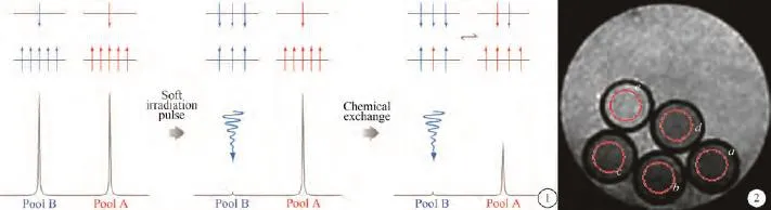
图1 化学交换饱和转移的二池模型 图2 4个pH值不同的离心试管(a~d)与装有等量水的试管(e)的磁化传递成像,施加了预报和脉冲,红圆圈所标定的是感兴趣区,测定结果a~d各试管信号强度不同Fig. 1 Model of Two pools for CEST Fig. 2 MT imaging of four centrifuge tubes with different pH values(a—d) and a tube w ith water (e) obtained w ith saturation pulse. The red circles denote regions of interest with different signal intensities for tubes a—d.
[References]
[1]Zhou J, Payen JF, W ilson DA, et al. Using the am ide proton signals of intracellular proteins and peptides to detect pH effects in MRI. Nature Medicine, 2003, 9(8): 1085-1090.
[2]Sun PZ, Zhou J, Sun W, et al. Suppression of lipid artifacts in amide p roton transfer imaging. M agnetic Resonance in Medicine, 2005, 54(1): 222-225.
[3]W en Z, Hu S, Huang F, et al. MR im aging of high-grade brain tumors using endogenous protein and peptide-based contrast. Neuro Image, 2010, 51(2): 616-622.
[4]Wei MB, Shen ZW, Xiao G, et al. Study of magnetic resonance im aging at 1.5 Tesla based on pH-sensitive m agnetization transfer technolog. Chin J Magn Reson Imaging, 2012, 3(1): 40-43.
韦茂彬, 沈智威, 肖刚, 等. 基于pH值敏感的磁化传递技术在1.5 T磁共振成像上的研究. 磁共振成像, 2012, 3(1): 40-43.
[5]W olff SD, Balaban RS. NMR imaging of labile p roton exchange. Journal of Magnetic Resonance, 1990, 86(1): 164-169.
[6]W ard KM, A letras AH, Balaban RS. A new class of contrast agents for MRI based on proton chemical exchange dependent saturation transfer (CEST). Journal of Magnetic Resonance, 2000, 143(1): 79-87.
[7]Wolff SD, Balaban RS. Magnetization transfer contrast (MTC) and tissue water proton relaxation in vivo. Magnetic Resonance in Medicine, 1989, 10(1): 135-144.
[8]Zhang TJ, Gong QY. Current status of magnetization transfer imaging in psychiatric disorders. Chin J Magn Reson Imaging,2010, 1(6): 468-472.
张体江, 龚启勇. 磁化传递成像及其在精神疾病中的研究现状. 磁共振成像, 2010, 1(6): 468-472.
[9]Zaiss M, Bachert P. Chemical exchange saturation transfer (CEST) and MR Z-spectroscopy in vivo: a review of theoretical approaches and m ethods. Physics in M edicine and Biology, 2013, 58(22): 221-269.
[10]Wu R, Xiao G, Zhou IY, et al. Quantitative chemical exchange saturation transfer(qCEST) MRI - omega plot analysis of RF-spillover-corrected inverse CEST ratio asymm etry for simultaneous determination of labile proton ratio and exchange rate. NMR in Biomedicine, 2015, 28(3): 376-383.
[11]Shen ZW, W ang LH, Dai ZZ, et al. Chem ical exchange saturation transfer (CEST)im aging of pH. Neuroscience and Biomedical Engineering, 2013, 1(2): 111-115.
[12]Wu RH, Shen ZW, Ning LB, et al. Magnetic resonance pH imaging. International Journal of Medical Radiology, 2010, 33(5): 409-410.
吴仁华, 沈智威, 宁立波, 等. 磁共振pH成像的研究. 国际医学放射学杂志, 2010, 33(5): 409-410.
[13]N ing LB, Qiu QC, X iao YU, et al. Chenm ical ex change saturation transfer and its app lication on MR pH imaging. Zhonghua Fang She Xue Za Zhi, 2011, 45(4): 414-416.
宁立波, 邱庆春, 肖叶玉, 等. 化学交换饱和转移在MR pH成像方面的应用. 中华放射学杂志, 2011, 45(4): 414-416.
[14]Zhou JY, Hong XH. Molecular imaging using endogenous cellular protein. Chinese Journal of Magnetic Resonance, 2013, 30(3): 307-321.
周进元, 洪晓华. 内源性蛋白质分子成像的研究进展. 波谱学杂志, 2013, 30(3): 307-321.
[15]Teng GJ, Cui Y. Research progress in molecular magnetic resonance imaging. Chin J Magn Reson Imaging, 2014, 5(S1): 31-36.
滕皋军, 崔莹. 磁共振分子影像学研究进展. 磁共振成像, 2014, 5(S1): 31-36.
[16]Kogan F, Hariharan H, Reddy R. Chem ical exchange saturation transfer (CEST)imaging: description of technique and potential clinical applications. Current Radiology Reports, 2013, 1(2): 102-114.
[17]Zhang T, Jia YL, Nie TT, et al. Study of a new method for imaging of GABA based on chemical exchange saturation transfer using a 7.0 T MR scanner. Chin J M agn Reson Imaging, 2015, 6(5): 385-389.
章桃, 贾岩龙, 聂婷婷, 等. 7.0 T MR γ-氨基丁酸化学交换饱和转移成像的新技术研究. 磁共振成像, 2015, 6(5): 385-389.
[18]Aime S, Carrera C, Delli Castelli D, et al. Tunable imaging of cells labeled w ith MRI-PARACEST agents. Angewandte Chemie, 2005, 44(12): 1813-1815.
[19]Srivastava AK, Kadayakkara DK, Bar-Shir A, et al. Advances in using MRI probes and sensors for in vivo cell tracking as app lied to regenerative medicine. Disease M odels & Mechanisms, 2015, 8(4): 323-336.
[20]Gilad AA, M cMahon MT, Walczak P, et al. Artificial reporter gene providing MRI contrast based on proton exchange. Nature Biotechnology, 2007, 25(2): 217-219.
[21]Xiao H, X iao G, Wu RH. M odeling and simulation of chem ical exchange saturation transfer m agnetic resonance im aging. Journal of Lanzhou University of Technology, 2014, 40(4): 71-75.
肖红, 肖刚, 吴仁华. 化学交换饱和传递磁共振成像建模与仿真. 兰州理工大学学报, 2014, 40(4): 71-75.
[22]Zhou J, Hong X, Zhao X, et al. APT-weighted and NOE-weighted image contrasts in glioma w ith different RF saturation powers based on magnetization transfer ratio asymmetry analyses. M agnetic Resonance in Medicine, 2013, 70(2): 320-327.
[23]Zaiss M, W indschuh J, Paech D, et al. Relaxation-compensated CEST-MRI of the human brain at 7 T: Unbiased insight into NOE and am ide signal changes in human gliob lastoma. NeuroImage, 2015, 112: 180-188.
[24]Yu H, Wang XL, Jiang SS, et al. A prelim inary study on identification of the brain metastatic tumors and the high-grade neuroepithelial tumors w ith am ide proton transfer m agnetic resonance im aging. Chinese Journal of Neurosurgery, 2015, 31(10): 1042-1046.
于昊, 王显龙, 蒋山姗, 等. 磁共振氨基质子转移成像鉴别脑转移瘤与高级别神经上皮肿瘤的初步探讨. 中华神经外科杂志, 2015, 31(10): 1042-1046.
[25]Ma B, Blakeley JO, Hong X, et al. Applying amide proton transfer-w eighted MRI to distinguish pseudoprogression from true progression in m alignant gliomas. J M agn Reson Imaging, 2016 Jan 20. doi: 10.1002/jm ri.25159.
[26]Sun PZ, Benner T, Kumar A, et al. Investigation of optim izing and translating pH-sensitive pulsed-chem ical exchange saturation transfer (CEST) imaging to a 3 T clinical scanner. Magnetic Resonance in Medicine, 2008, 60(4): 834-841.
[27]Cao Z, Yang ZX, Zhou ZH, et al. MR pH imaging. In Book: Edited by Lang ZJ, M iao YW, W u RH, et al. MR new techniques. Shanghai: Shanghai Sci Tech Publisher, 2015: 192-194.
曹震, 杨忠现, 赵芝弘, 等. MR酸碱度成像. 郎志谨, 苗延巍,吴仁华, 等. MRI 新技术, 上海: 上海科学技术出版社, 2015: 192-194.
[28]Wang R, Li SY, Chen M, et al. Amide proton transfer magnetic resonance im aging of A lzheimer's d isease at 3.0 Tesla: a prelim inary study. Chinese Medical Journal, 2015, 128(5): 615-619.
[29]Wang L, Li CM, Zhang C, et al. A preliminary study on amide proton transfer MR imaging at 3.0 T of the substantia nigra and red nucleusinParkinsondisease. Zhonghua Fang She Xue Za Zhi, 2015, 49(2): 81-84.
王蕊, 李春媚, 张晨, 等. 帕金森病患者黑质和红核的3.0 T M R氨基质子转移成像初步研究. 中华放射学杂志, 2015, 49(2): 81-84.
[30]Chen SZ, Yuan J, Deng M, et al. Chemical exchange saturation transfer (CEST)MR technique for in-vivo liver imaging at 3.0 tesla. Eur Radiol, 2015 Sep 3.[Epub ahead of print].
[31]Forsen S, Hoffman RA. Study of moderately rapid chemical exchange reactions by m eans of nuclear m agnetic double resonance. The Journal of Chem ical Physics, 1963, 39(11): 2892-2901.
[32]Guivel-Scharen V, Sinnwell T, Wolff SD, et al. Detection of proton chemical exchange between metabolites and water in biological tissues. Journal of Magnetic Resonance, 1998, 133(1): 36-45.
[33]Ning LB. pH imaging pulse sequence designin the clinical m agnetic resonance scannerbased on chem ical exchange saturation transfer. Shantou: Shantou University, 2010.
宁立波. 基于化学交换饱和转移机制的磁共振pH成像脉冲序列设计. 汕头: 汕头大学, 2010.
[34]Liu TZ. Study on detection of intracranial tumors with magnetization transfer sequence. Shantou: Shantou University, 2011.
刘天柱. 应用磁化传递序列检测颅内肿瘤的研究. 汕头: 汕头大学, 2011.
[35]Lin TF. Chem ical exchange saturation transfer imaging of hyperacute brain infarction using 1.5 T scanner. Shantou: Shantou University, 2011.
林泰锋. 超急性期脑梗塞1.5 T磁共振化学交换饱和转移成像.汕头: 汕头大学, 2011.
[36]Sherry AD, Woods M. Chemical exchange saturation transfer contrast agents for magnetic resonance imaging. Annual Review of Biomedical Engineering, 2008, 10(10): 391-411.
[37]Desmond KL, Stanisz GJ. Understanding quantitative pulsed CEST in the presence of MT. Magnetic Resonance in Medicine, 2012, 67(4): 979-990.
[38]W oods M, Pasha A, Zhao P, et al. Investigations into whole water, prototropic and am ide proton exchange in lanthanide(III) DOTA-tetraamide chelates. Dalton Transactions, 2011, 40(25): 6759-6764.
[39]Sun PZ, Longo DL, Hu W, et al. Quantification of iopamidol multi-site chemical exchange properties for ratiometric chemical exchange saturation transfer (CEST) imaging of pH. Physics in Medicine and Biology, 2014, 59(16): 4493-4504.
[40]Zhou YL, Li HL. Applications of nuclear overhauser effect. Orgnic Chem istry, 1986(1): 1-18.
周原朗, 李华立. 核OVERHAUSER效应(NOE)的应用. 有机化学, 1986(1): 1-18.
[41]Dai Z, Ji J, Xiao G, et al. Magnetization transfer prepared gradient echo MRI for CEST imaging. PloS One, 2014, 9(11): 112219.
[42]Lin Z, Oostenbrink C, van Gunsteren WF. On the use of onestep perturbation to investigate the dependence of NOE-derived atom-atom distance bound violations o f pep tides upon a variation of force-field param eters. European Biophysics Journal, 2014, 43(2-3): 113-119.
[43]Zaiss M, W indschuh J, Goerke S, et al. Dow nfield-NOE-suppressed am ide-CEST-MRI at 7 Tesla provides a unique contrast in human gliob lastoma. Magnetic Resonance in Medicine, 2016, in press. DOI: 10.1002/m rm.26100.
[44]Sun PZ, Farrar CT, Sorensen AG. Correction for artifacts induced by B(0) and B(1) field inhomogeneities in pH-sensitive chemical exchange saturation transfer (CEST) imaging. Magnetic Resonance in M edicine, 2007, 58(6): 1207-1215.
[45]Cheng XF, W u RH. MR-based methods for pH measurement in brain tumors: current status and clinical potential. Journal of Gannan University, 2011, 31(4): 506-509.
成小芳, 吴仁华. 基于磁共振技术的脑肿瘤pH检测方法的现状及临床应用前景. 赣南医学院学报, 2011, 31(4): 506-509.
[46]Cheng XF, Wu RH. MR-based methods for pH measurement in brain tumors: current status and clinical potential. In Book: Edited by Ana L. Abujam ra: Brain Tumors, 2011, in Tech 287-302.
[47]Sun PZ, Xiao G, Zhou IY, et al. A method for accurate pH mapping w ith chemical exchange saturation transfer (CEST) MRI. Contrast M edia & M olecular Imaging, 2016, in press. DOI: 10.1002/cmmi.1680.
[48]Sun PZ, Wang Y, Dai Z, et al. Quantitative chemical exchange saturation transfer(qCEST) MRI--RF spillover effect-corrected omega plot for simultaneous determination of labile proton fraction ratio and exchange rate. Contrast Media & Molecular Imaging, 2014, 9(4): 268-275.
[49]Longo DL, Sun PZ, Consolino L, et al. A general MRI-CEST ratiometric approach for pH imaging: demonstration of in vivo pH m apping w ith iobitridol. Journal of the American Chem ical Society, 2014, 136(41): 14333-14336.
[50]M cVicar N, Li AX, Suchy M, et al. Sim ultaneous in vivo pH and temperature mapping using a PARACEST-MRI contrast agent. M agnetic Resonance in M edicine, 2013, 70(4): 1016-1025.
[51]Bodet O, Goerke S, Behl NG, et al. Am ide proton transfer of carnosine in aqueous solution studied in vitro by WEX and CEST experiments. NMR in Biomedicine, 2015, 28(9): 1097-1103.
[52]Tietze A, Blicher J, M ikkelsen IK, et al. Assessment of ischemic penumbra in patients w ith hyperacute stroke using amide proton transfer (APT) chemical exchange saturation transfer (CEST) MRI. NMR in Biomedicine, 2014, 27(2): 163-174.
[53]Sun PZ, Cheung JS, Wang E, et al. Association between pH-w eighted endogenous am ide p ro ton chem ical ex change saturation transfer MRI and tissue lactic acidosis during acute ischem ic stroke. Jou rnal of Cereb ral B lood Flow and Metabolism, 2011, 31(8): 1743-1750.
[54]Huang D, Li S, Dai Z, et al. Novel gradient echo sequencebased am ide p roton transfer m agnetic resonance im aging in hyperacute cerebral infarction. M olecular M edicine Reports, 2015, 11(5): 3279-3284.
[55]Wang M, Hong X, Chang CF, et al. Simultaneous detection and separation of hyperacute intracerebral hemorrhage and cerebral ischemia using amide proton transfer MRI. Magnetic Resonance in Medicine, 2015, 2(19): 1608-1612.
[56]Lee DH, Heo HY, Zhang K, et al. Quantitative assessment of the effects of water proton concentration and w ater T changes on am ide p roton transfer (APT) and nuclear overhauser enhancement (NOE) MRI: The origin of the APT imaging signal in brain tumor. Magnetic Resonance in Medicine, 2016, in press. DOI: 10.1002/m rm.26131.
[57]Wu KW, W u RH, Zhang MM. Preliminary study of breast cancer in magnetic resonance im aging based on am ide proton transfer. Functional and M olecular Medical Imaging, 2013, 2(1): 35-39.
伍康伟, 吴仁华, 张苗苗. 基于氨基转移机制的乳腺癌磁共振成像初步研究. 功能与分-r医学影像学(电子版), 2013, 2(1): 35-39.
[58]Dula AN, Dewey BE, Arlinghaus LR, et al. Optim ization of 7 T chem ical exchange saturation transfer param eters for validation o f g lycosam inog lycan and am ide proton transfer of fibroglandular breast tissue. Radiology, 2015, 275(1): 255-261.
M agnetic resonance am ide p roton transfer (APT) imaging: description of technical principles and potential applications
WU Ren-hua*
Department of M edical Imaging, the 2nd A ffiliated Hospital, Shantou University M edical College, Shantou 515041, China
Am ide proton transfer (APT) imaging is related to chem ical exchange saturation transfer (CEST) technique. This review focuses on description of APT technical principles, factors influencing APT imaging, and relationship between in vivo APT imaging and nuc lear Overhauser effect (NOE). Potential clinical applications of APT technique, such as pH imaging, temperature imaging, as well as APT-weighted imaging for organs and tissues, are discussed.
Am ide proton transfer; Chem ical exchange saturation transfer; Nuclear Overhauser effect; pH imaging; Tem perature imaging
国家自然科学基金项目(编号:81471730)
汕头大学医学院第二附属医院,汕头515041
ail: cjr.wurenhua@vip.163.com
2016-02-17
接受日期:2016-04-03
R445.2
A
10.12015/issn.1674-8034.2016.04.003
吴仁华. 磁共振氨基质子转移成像技术原理和应用. 磁共振成像, 2016, 7(4): 254–258.
*Correspondence to: Wu RH, E-mail: cjr.wurenhua@vip.163.com
猜你喜欢
现代畜牧科技(2021年9期)2021-10-13
国际放射医学核医学杂志(2020年4期)2020-07-27
新农业(2018年3期)2018-07-08
新农业(2018年3期)2018-07-08
高中数理化(2016年19期)2016-11-14
电源技术(2016年9期)2016-02-27
西北工业大学学报(2015年4期)2016-01-19
新疆大学学报(自然科学版)(中英文)(2014年2期)2014-11-06
太空探索(2014年6期)2014-07-10
江西理工大学学报(2013年1期)2013-03-20